Early detection of melanoma and assessment of asymptomatic people at high risk

Lesion thickness is the strongest predictor of prognosis in patients with a primary cutaneous melanoma. Therefore, identifying and investigating suspicious lesions at the earliest possible clinical stage can improve patient outcomes. Furthermore, identifying patients at high risk of melanoma is essential to target those who will benefit most from increased surveillance and education on sun smart behaviour.

1 comments
save
share
feedback
Log in

Published: 15 December 2021


Key practice points:

  • Encourage patients to report any abnormal naevi or other skin lesions and use any appropriate opportunity to assess a patient for suspicious lesions, e.g. during clinical examination for another reason. Survival is inversely correlated with lesion thickness; therefore, early detection is key to improving survival outcomes.
  • After identifying a lesion of concern, assess the likelihood of melanoma. Consider history of change, characteristics of the lesion including any itching, bleeding or pain, family history or other risk factors; use the ABCDEFG checklist in conjunction with dermatoscopy (using the Chaos and Clues method of pattern analysis) to examine the lesion. Depending on the patient’s risk of melanoma, the rest of the body may need to be checked for other suspicious lesions.
  • A narrow complete excisional biopsy with 2 mm margins should be performed on suspicious lesions; otherwise, refer the patient to a dermatologist or surgeon for assessment or removal. Low concern flat lesions can be monitored using digital dermatoscopy (clinical and dermatoscopic photographs) over three months and then as required, however, undiagnosed raised lesions (nodules) should be excised rather than monitored.
  • Evaluate patients who have risk factors for melanoma to determine the most appropriate interval for whole body skin examination. Patients at very high risk, e.g. family and personal history of melanoma and/or multiple atypical naevi, are recommended to undergo long-term surveillance with total body photography.
  • Encourage all patients at increased risk of melanoma to regularly examine their skin, document any changes over time and return for re-examination if changes are observed or new lesions appear

For general information on melanoma risk factors and prevention strategies, see: https://bpac.org.nz/2020/melanoma-part1.aspx

The majority of melanomas develop from uncontrolled melanocyte proliferation within the epidermis (melanoma in situ), which can then spread to the dermis (invasive melanoma) and in some cases, to regional lymph nodes and other tissues and organs (metastatic melanoma).1, 2 Less commonly, melanomas may arise within the dermis, therefore, do not have an in situ phase. In very rare cases, melanomas can originate in internal tissues, such as the brain and the eye.2

Melanoma lesion thickness is the strongest predictor of prognosis. In general, the thinner the lesion, the better the outcome for the patient; people with melanomas ≤ 1 mm thick have a ten-year survival rate of up to 92% compared with only 50% when melanomas are > 4 mm thick.3

Most melanomas are first recognised by the person themselves or family members, however, there is evidence that those identified by health professionals are thinner and have a more favourable prognosis.3, 4 Therefore, opportunistic skin checks are valuable; be suspicious of lesions with atypical features or a history of change.5

Subtypes of melanoma

There are four main clinical subtypes of melanoma, all of which have the potential to metastasise if they invade the dermis (Table 1).1, 2 These include superficial spreading melanoma, nodular melanoma, lentigo maligna melanoma and acral lentiginous melanoma. Less common subtypes include desmoplastic and mucosal lentiginous melanoma.1, 2

Prognosis differs based on melanoma subtype; nodular and acral lentiginous melanomas are usually thicker at diagnosis and associated with a lower five-year survival rate compared with superficial spreading and lentigo maligna melanomas.4


Table 1: The four main clinical subtypes of melanoma (images supplied by DermNet NZ).1, 2, 4

Type Description Example image based on visual inspection Example image based on dermatoscopic inspection
Superficial spreading melanoma

The most common type of melanoma. Often has a prolonged pre-invasive in situ phase, growing slowly over months to decades. Generally found on the trunk and associated with a history of sunburn and the presence of large numbers of melanocytic naevi. Begins as a flat pigmented patch of irregular shape with variable border abruptness and increasing colours as it deepens.

Dermatoscopic clues* may include blue/grey structures, eccentric structureless areas, multiple irregular brown dots or clods, thick lines, radial lines.

Nodular melanoma

The second most common type of melanoma. Characterised by a rapidly growing (over several weeks to months), pink, red, brown or black nodule. The pigmentation within nodular melanomas is often more uniform than in superficial spreading forms. Nodular melanomas may be more likely to bleed or ulcerate than superficial spreading melanomas and do not have an in situ radial growth phase.

Dermatoscopic clues* may include blue/grey structures, eccentric structureless areas, white structures and polymorphous vessels.

Lentigo maligna (head and neck) and lentiginous melanoma (upper trunk)

Found on sun-damaged skin in older people. Lesions typically have a long pre-invasive in situ stage (years to decades). Characterised by a slowly enlarging, irregularly pigmented patch.

Dermatoscopic clues* may include asymmetrical, follicular pigmentation (on the face) and polygons.

Acral lentiginous melanoma

Found on the palms of the hands and soles of the feet; may involve finger or toenails. Acral lentiginous melanomas account for fewer than 5% of all melanomas in New Zealand. This form is the most common type of melanoma in people with dark skin, including Māori and Pacific peoples. There is a higher rate of amelanotic melanoma among acral lesions.

Dermatoscopic clues* may include parallel lines on ridges. Melanoma arising from a nail matrix causes irregular expanding longitudinal bands on the nail plate.

*See: “Dermatoscopy should be an essential skill in primary care - Section: Methods and interpretation of dermatoscopy)” for specific information on the Chaos and Clues dermatoscopic method of revised pattern analysis

For further reading on the subtypes of melanoma using the Chaos and Clues dermatoscopic method of pattern analysis, see: melnet.org.nz/uploads/Revised-Pattern-Analysis-and-Chaos-and-Clues.pdf and dermnetnz.org/assets/Uploads/Chaos-and-Clues-Landscape-reduced-ilovepdf-compressed.pdf

Look for the “ugly duckling"

Melanomas exhibit significant heterogeneity and can sometimes be difficult to identify clinically.1 In many cases the “ugly duckling” principle will prompt the initial suspicion of melanoma, with the rationale being that any lesion that is dissimilar in appearance, e.g. size, colouring and shape, to other moles, freckles or lesions, should undergo further scrutiny (see: Figure 1 and “Clinical checklists in conjunction with dermatoscopy can support the diagnostic suspicion of melanoma”). This may be a particularly useful approach in patients with large numbers of solar lentigos, seborrhoeic keratoses or melanocytic naevi (see: “Glossary of terms”).

Evaluating the lesion of concern in the context of other lesions enables an understanding of what is considered “normal” for the patient. It is best practice that a lesion is examined with dermatoscopy, rather than visual inspection alone (see: “Dermatoscopy should be considered an essential skill in primary care”).5 Ideally after identifying a suspicious lesion, examine the rest of the body for other lesions of concern.


Figure 1. Example of an “ugly duckling” lesion (image supplied by DermNet NZ).

After assessing the patient’s lesion of concern in the context of other lesions, ask about factors such as history of change, characteristics of the lesion including itching, bleeding or pain and symptom duration, family history and other risk factors, e.g. age, significant sun exposure, light complexion (Table 2), and work through the ABCDEFG checklist in conjunction with dermatoscopy to determine the likelihood of melanoma (see: “Dermatoscopy should be an essential skill in primary care - Section: Methods and interpretation of dermatoscopy)”.2, 5

An updated and expanded ABCDEFG checklist helps to identify all types of melanoma lesions, including nodular and thick melanomas which were not always recognised early using the traditional ABCDE criteria.6

The ABCDEFG checklist:5, 7

  • Asymmetry – one half of the lesion does not match the other
  • Border irregularity – notched, blurred, ragged and especially variable edges
  • Colour variegation – different colours such as brown, black, white, red or blue within the same lesion
  • Different – the lesion looks different from other spots, freckles or moles (i.e. an “ugly duckling”)
  • Evolution or elevation – any change in a lesion over time is suspicious especially if documented by digital dermatoscopy
  • Firm – the lesion is firm to the touch
  • Growing – the majority of melanoma are more than 6 mm in diameter and keep growing

Bleeding, crusting, inflammation, pruritus and pain are not included in the ABCDEFG checklist, but are additional features that may be present, particularly with nodular and thick melanomas. The ABCDEFG checklist criteria are not unique to melanomas and some early-stage melanomas may not initially display any of these characteristics. Any lesion that continues to grow or change in size, shape, colour or elevation over a period of more than one month is suspicious of melanoma.4,8 However, seborrhoeic keratoses (which have many common features with melanoma based on the ABCDEFG checklist) and inflammatory disorders should also be considered.

If a high concern lesion is identified, check the rest of the body for other suspicious lesions

For further information on the ABCDEFG criteria, see: https://dermnetnz.org/topics/abcdes-of-melanoma

If your practice does not have a dermatoscope and there is clinical concern based on visual inspection and history, consider excision biopsy or refer the patient for further assessment if there is timely access to an appropriate service in your region.

Other diagnostic tools may be considered, as appropriate

Serial digital photography. Digital clinical photographs of low-concern lesions can be taken either by the patient themself or a health professional at regular intervals to monitor changes over time.4 Clearly label photographs taken at your practice, e.g. document the date, body site, lesion size and any other notable characteristics, and consider privacy issues in terms of where in the patient record the photos are saved and who has access to them. The addition of dermatoscopy images significantly enhances follow-up.

Total body photography. This is the use of clinical photography (ranging between 12 and 24 baseline photographs) to provide a record of a patient’s entire skin surface, rather than photographs of individual lesions alone.4 Consider how these photographs are to be documented and any potential privacy concerns (as above). Total body photography is most useful for detecting new lesions and for assessing changes in pigmented lesions over time. There are dedicated services, e.g. MoleMap NZ, that provide total body photography with review by accredited dermatologists, although cost may be a barrier for many people.

Ophthalmoscopy. Observation using an ophthalmoscope can help identify choroidal melanomas which are most commonly orange pigmented, dome-shaped and may be associated with an exudative retinal detachment.12

Smartphone apps. Validated smartphone apps may be used to aid early skin cancer detection by enabling people to archive and share photos of their naevi with a skin specialist or dermatologist. This approach may help to overcome reluctance that some people have in seeking medical advice about their skin lesions, however, may provide false reassurance and potentially delay diagnosis; studies are ongoing to assess these risks.13 Smartphone apps do not replace a skin examination by a clinician and cost may be a barrier for some people.

For further information on smartphone skin check apps, see: https://healthify.nz/apps/s/skin-check-apps/

Artificial intelligence. An emerging technology assisting in the diagnosis of suspicious skin lesions where set algorithms automatically categorise lesions into diagnostic classes, e.g. naevus, benign, malignant or melanoma.14 Validated tools can be used alongside expert dermatoscopic analysis to enhance clinical best practice.5

For early stage melanoma, timely intervention is particularly important as the overall survival rate is close to 100% with prompt surgical excision, which aims to eliminate all localised abnormal cells, i.e. it is essentially “curative”.1 For patients with advanced or stage IV melanoma with regional or more widespread metastases, treatment options are likely to include surgery, targeted therapy or immunotherapy and less commonly, radiation or chemotherapy, under the guidance of secondary care and are associated with widely variable outcomes.

Excisional biopsy is required for high concern lesions

The New Zealand Melanoma Quality Statements instruct that a narrow complete excisional biopsy with 2 mm margins and of sufficient depth to avoid transection at the base should be performed under local anaesthesia on any pigmented lesion suspicious of melanoma generally within two weeks of being identified (see: ‘Melanoma and excision terminology’ in “Glossary of terms” and “Criteria for a high concern lesion”).4, 5 Depending on the size, body site, ease of obtaining clear margins and the suspected type of lesion, this may be performed in primary care. If direct closure with a 2 mm margin is not possible or if the excision cannot be performed in general practice for any reason, e.g. if there is concern of lymph node involvement, urgently refer to a dermatologist or surgeon.1 In some regions, funded referral to a general practitioner with a special interest (GPSI) in skin cancer may be available. Partial biopsies, e.g. punch and shave biopsies, should generally only be performed by melanoma specialists and in certain clinical situations, e.g. if the lesion is unusually large or for some acral lesions.2

If melanoma is diagnosed, the pathology report will include:2

  • Tumour thickness (Breslow thickness) to the nearest 0.1 mm from the top of the granular layer to the deepest malignant cell (for invasive melanoma only)
  • Clark level of invasion
    • Level 1 – melanoma in situ (confined to the epidermis)
    • Level 2 – melanoma invades the papillary dermis
    • Level 3 – melanoma fills the papillary dermis
    • Level 4 – melanoma invades the reticular dermis
    • Level 5 – melanoma invades subcutaneous tissue
  • Clearance margins
  • Mitotic rate (how fast cells are proliferating)
  • Presence or absence of ulceration

This information can then be used to direct subsequent management decisions, which will involve either wide local excision of the melanoma based on Breslow thickness in primary care or referral to secondary care (see: “Glossary of terms”). For details on appropriate margin sizes for wide local excision, see “Quality statement 5.1” in www.melnet.org.nz/uploads/Melanoma-Quality-Statements-FINAL-published-November-2021.pdf

Best practice tip: Histological diagnosis of melanoma by a pathologist is often difficult. Ensure the clinical request form accompanying the specimen includes the following information: history, specimen site, biopsy type and clinical/dermatoscopic description of the lesion.5 Some pathologists find annotated clinical and dermatoscopic photographs useful, particularly for borderline lesions with attention drawn to areas of specific concern, e.g. eccentric pigmentation, as melanoma might be arising within a melanocytic naevus. “Derm dotting”* can be used to mark the area of concern by applying coloured nail polish to the areas where there are concerning features.5

*“Derm dotting” is a technique where a spot of coloured nail polish, e.g. red, orange, blue or black, is applied using a toothpick or fine brush on the area of concern at least 30 seconds prior to immersing in formalin.5, 16 The nail polish does not affect the tissue morphology and helps the pathologist to identify the specific concerning feature(s) when examining the specimen under a microscope to make a more accurate diagnosis.5, 16

Keep an eye on low concern flat lesions

A low concern lesion describes those with atypical features but without evidence of melanoma.4, 5 Observation over three months* (using digital dermatoscopy) is most appropriate for low concern, flat (≤ 1 mm), pigmented lesions, and then as required.4, 5 If there are known barriers to reattendance, consider a lower threshold for excision.17 Monitoring alone is not an appropriate strategy for patients with thickened melanocytic lesions (> 1 mm); if the diagnosis is unclear, excise the lesion or refer the patient to a dermatologist.4

*A surveillance period of six months is recommended if there is suspicion of lentigo maligna4

Factors such as older age, sun damage, personal or family history of melanoma, multiple naevi (e.g. > 100) or actinic keratoses, place people at higher risk of developing melanoma (Table 2).5 Evaluate those with risk factors for melanoma to determine the most appropriate interval for whole body skin examination. Validated models (see link below) are available that integrate patient risk factors to produce an overall risk level for developing melanoma.5

High risk patients: It is currently recommended that patients with ≥ 2 first-degree relatives with a history of melanoma when aged < 40 years and with a personal history of melanoma and/or multiple atypical naevi be placed under long-term skin surveillance, ideally with a clinician experienced in dermatoscopy.5 Visual examination in conjunction with dermatoscopy and total body photography should occur every 12 months to detect new lesions or track changes in existing lesions.5 Also consider referral to regional clinical genetics services for further assessment.5

All patients: Encourage all patients at increased risk of melanoma to:4, 5

  • Follow sun smart principles, e.g. “slip, slop, slap and wrap”. A smartphone app, e.g. UVLens, may be used to monitor daily UV index levels. For further information on being sun smart, see: www.sunsmart.org.nz
  • Regularly examine their skin (including areas not normally exposed to the sun, e.g. soles of feet, between toes, under nails, genitalia and ask a relative or friend to check difficult to see areas) and document changes over time. Patients can use the ABCDEFG checklist to aid with self-checks; alternatively, a simpler acronym such as SCAN (sore, changing, abnormal, new) may be easier for the patient to remember.
  • Return to primary care if changes are observed or new lesions appear; emphasise that smartphone apps and self-skin checks should not replace skin examination by a clinician

There are no validated New Zealand specific tools for calculating a patient’s risk of developing melanoma. For an example of an Australian tool, see: www.melanomarisk.org.au/

N.B. People living in New Zealand should enter ‘Tasmania’ as the ‘Region of Australia most lived in’ to receive the most appropriate risk profile.5

Table 2. Risk factors for melanoma.2, 4–6, 18, 19

Risk factor Explanation

Age

The risk of melanoma increases with age, likely due to an increased probability of intermittent UV overexposure events and immune suppressive effects of ageing. However, younger people are still considered a priority for prevention advice due to sun-seeking behaviours and increased participation in outdoor activities. Approximately 19% of melanomas arise in people aged < 50 years.

Sex/gender

Overall, males have a higher risk of melanoma than females, other than those aged ≤ 50 years. Males are more likely to have lesions on their trunk, while females are more likely to have lesions on their legs. Differences are presumably due to a combination of behavioural and biological factors.

Personal or family history of melanoma

A number of genetic mutations, e.g. CDKN2A, increase the risk of developing melanoma. People who have a first-degree family member with a history of melanoma are twice as likely to develop melanoma than those without. A personal history of melanoma increases the risk of new melanoma ten times, and a personal history of non-melanoma skin cancer doubles the risk compared to those without a history of skin cancer.

Eye/hair colour

In general, people with blue/grey eyes or blonde/red hair have a higher risk of melanoma than people with brown eyes and dark hair

Skin colour/type
(before tanning)

The risk of skin cancer is influenced by the amount of melanin produced by melanocytes, more melanin results in greater protection against UV radiation. Therefore, people with darker skin types* have a considerably lower risk of melanoma compared to those with “fair” skin or skin that burns easily.

The Fitzpatrick skin type system is one method of identifying people requiring increased sun protection based on their level of melanin pigmentation. With this system, skin types I and II are considered to have a high risk of sun damage:

  • Type I – pale white skin, blue/green eyes, blonde/red hair – always burns, does not tan
  • Type II – fair skin, blue eyes – burns easily, tans poorly
  • Type III – darker white skin – tans after initial burn
  • Type IV – light brown skin – burns minimally, tans easily
  • Type V – brown skin – rarely burns, tans darkly easily
  • Type VI – dark brown or black skin – never burns, always tans darkly

Immune suppression

A weakened immune system can increase the risk of melanoma, e.g. due to HIV, leukaemia or immunosuppressant medicine use, e.g. after organ transplantation

Number of moles

Approximately one-third of melanomas develop within pre-existing melanocytic naevi. Both genetic factors and UV exposure influence the development of naevi. A high number of naevi (> 100) and large/atypical naevi (> 5) increase the risk of melanoma.

Type of moles

Geographic location

Some locations in New Zealand have higher reported rates of melanoma than others. The association of geographic location and melanoma risk is complicated and may be due to factors such as ethnic distribution, early diagnosis, reporting practices, atmospheric factors, outdoor occupation and sun-seeking behaviours.

Ultraviolet radiation exposure

  • History of sunburn
  • Intermittent UV overexposure
  • Outdoor employment/leisure
  • Sunbed use

UV radiation from the sun (and tanning devices) is the primary cause of the uncontrolled melanocyte proliferation seen in melanoma in white skin. New Zealand has relatively high levels of UV radiation compared to the same latitudes in the Northern hemisphere. Given that multiple factors relating to UV exposure are modifiable, these represent a core focus of prevention strategies. Consider recommending the use of smartphone apps, e.g. UVLens, which can forecast UV index levels. Some apps, including UVLens, personalise index levels according to skin type, geographic location, time of day and year and cloud cover.

Non-modifiable    Partly or potentially modifiable    Modifiable

* There are no systematic investigations linking ethnicity to skin type in New Zealand, and Māori and Pacific peoples, as with all ethnic groups, have heterogeneity in skin type.3 However, at a population level there are obvious differences, with melanoma rates being over five times higher in those of European descent compared with Māori and Pacific peoples. Additional genetic and behavioural factors beyond melanin production likely also play a role in melanoma risk for different ethnicities.


Early detection

  • Use any appropriate opportunity to examine a patient for suspicious lesions
  • If a suspicious lesion is identified, determine the likelihood of melanoma:
    • Consider history of change, characteristics of the lesion including itching, bleeding or pain and symptom duration, family history and other risk factors for melanoma, e.g. age, significant exposure to sunlight, fair complexion
    • Use the ABCDEFG checklist in conjunction with dermatoscopy (using the Chaos and Clues method of pattern analysis) to examine the lesion. If dermatoscopy is not available and there is clinical concern, excise the lesion or refer the patient for further assessment.
  • If the patient has a high concern lesion, check the rest of the body for other suspicious lesions
  • Perform a narrow complete excisional biopsy with 2 mm margins, and sufficient depth to avoid transection at the base, on high concern lesions, or refer the patient to a dermatologist or surgeon for assessment or removal
  • Monitor low concern flat lesions over three months using digital dermatoscopy and then as required. Advise patients to return if rapid changes are observed between appointments. Undiagnosed raised lesions (nodules) should be excised rather than monitored.

ABCDEFG checklist

  • Asymmetry
  • Border irregularity
  • Colour variegation
  • Different
  • Evolution or elevation
  • Firm
  • Growing

Chaos and Clues

Check for any evidence of chaos, i.e. more than one pattern, asymmetry of colour, structure and border abruptness, and if identified, look for one or more clues to malignancy, e.g.:

  • Eccentric structureless areas
  • Grey or blue structures
  • Large polygons
  • Parallel lines on ridges (palms and soles), chaotic lines (nails)
  • Segmental radial lines or pseudopods
  • Peripheral black dots or clods
  • Polymorphous vessels
  • Thick lines that are reticular or branched
  • White lines

Asymptomatic people at high risk

  • Evaluate patients with a history of melanoma or other skin cancers, for melanoma risk factors, e.g. multiple naevi or actinic keratoses, to determine the most appropriate interval for while body skin examination
  • Patients with ≥ 2 first-degree relatives with a history of melanoma when aged < 40 years and with a personal history of melanoma and/or multiple atypical naevi should be placed under long-term skin surveillance, ideally with a clinician experienced in dermatoscopy. A visual examination in conjunction with dermatoscopy and total body photography should occur every 12 months. Also consider referral to clinical genetics service.
  • Encourage all patients at increased risk of melanoma to follow sun smart behaviours, regularly examine their skin and document any changes over time. If changes are observed or new lesions appear, advise patients to return for re-examination; emphasise that smartphone apps should not replace a skin examination by a clinician.

Dermatoscopy

Dot: Structure too small to describe its shape

Chaos: More than one pattern in a pigmented lesion associated with asymmetry of colour, structure or border abruptness

Clod: Defined structure (i.e. not a line) that can be any shape or colour

Clues: Used as part of the Chaos and Clues dermatoscopic method of pattern analysis to determine whether a lesion should be excised. In a lesion displaying chaos, if there are one or more clues to malignancy, the lesion should be excised. Clues to malignancy in a pigmented lesion are: grey/blue structures, eccentric structureless areas, thick lines that are reticular or branched, peripheral black dots/clods, segmental radial lines or pseudopods, white lines, polymorphous vessels and large polygons. Parallel lines on ridges are a clue on palms and soles, and chaotic lines are a clue on nails.

Pattern: An arrangement of lines (most often reticular, but can be branched, parallel or radial), dots, clods or structureless zones

Pseudopod: A radial line with a bulbous end

Reticular pattern: A complete or incomplete network of intersecting lines

Revised pattern analysis: A system of describing the patterns and features of a pigmented lesion according to structures, colour and symmetry. A modification of the revised pattern analysis is called Chaos and Clues; revised pattern analysis of a lesion shows chaos, the specific features of the lesion reveal clues to malignancy.

Structureless zone: Lacks lines, dots or clods

Common benign skin lesions

Angioma: Benign vascular skin lesion, usually < 1 cm in diameter. Angiomas often appear black to the naked eye, however, dermatoscopic examination may show red, purple or blue clods.

Atypical naevus: Characterised by at least three of the following: size > 5 mm, ill-defined or blurred border, irregular margin resulting in an unusual shape, colour variation - mostly pink, tan, brown or black, flat and bumpy components. Atypical naevi often arise on the mid-upper back, breasts and genital areas. Dermatoscopy may show reticular, globular, homogenous or regression structures, an irregular vascular pattern and grey-blue areas.

Dermatofibroma: Benign fibrous nodule, often solitary and on the limbs. Most often a firm papule or nodule < 1.5 cm in diameter and may be pink or brown, often with a hypopigmented centre. Dermatofibromas are usually asymptomatic; however, they can also be painful, tender or itchy. The most common dermatoscopic feature is a whitish centre with fine radial lines or diffuse pigment fading peripherally.

Lentigo: Pigmented, flat or slightly raised lesion with a clearly defined border. May have a smooth or slightly dry surface. The solar lentigo is found on sun exposed sites. A solitary facial solar lentigo can at times be difficult to distinguish from melanoma in situ. Dermatoscopy shows a sharp, often scalloped border and light brown pigment with subtle structures.

Melanocytic naevus: Often referred to as a mole. Benign skin lesion of developmental origin; congenital or arises in childhood or early adult life within the dermis, at the junction of dermis and epidermis or both. Naevi vary widely in appearance and type. Dermatoscopy shows symmetry of patterns; on most body sites this can be structureless, reticular in a junctional naevus, aggravated clods in a dermal naevus or a combination of reticular and clod patterns in a compound naevus. Site-specific characteristics of junctional naevi include a pseudoreticular pattern in facial naevi and a parallel furrow pattern in acral naevi, i.e. palms, soles, fingers or toes.

Seborrhoeic keratosis: Benign keratinocytic proliferation often brown, black or tan but can also be pale, yellow or have several colours. Seborrhoeic keratoses vary widely in appearance, e.g. they can be solitary or multiple, flat or raised papules or plaques with a smooth, waxy or warty surface and range in diameter. As they are often irregular in structure and colour and change over time, they can be confused with melanoma. They commonly have a sharp border around the lesion that results in a stuck-on appearance. On sun exposed sites, a seborrhoeic keratosis may evolve within a solar lentigo. Dermatoscopy may show scattered irregular, superficial yellow, orange, grey, brown or blackish clods, scattered round or oval white or light-yellow clods (both best seen with non-polarised light) and thick curved lines forming a brain-like or seaweed pattern.

Pre-cancerous lesions and non-melanoma skin cancers

Actinic keratosis: Also known as solar keratosis. Actinic keratosis is a pre-cancerous keratinocytic (scaly) flat or thickened papule or plaque located on habitually sun exposed sites. They may be red or pigmented and patients may report tenderness or be asymptomatic. On facial sites, dermatoscopy shows prominent follicles in a red background, known as the strawberry pattern. Pigmented actinic keratosis also shows superficial angulated brown lines.

Basal cell carcinoma: Locally invasive keratinocytic skin cancer that varies in size, appearance and histology, but tends to have a rolled border. Often described as a slow growing, pink or pigmented shiny plaque or nodule that spontaneously bleeds or ulcerates (Figure 2). May invade deeply but rarely metastasises. Dermatoscopy on pigmented lesions may show blue ovoid structures, grey dots, leaf-like converging radial lines and polarised white structures on a shiny background of stroma (pale, pink, light brown or grey). Dermatoscopy on non-pigmented basal cell carcinomas may show polarised white structures and vessels are often irregular, linear or branched.

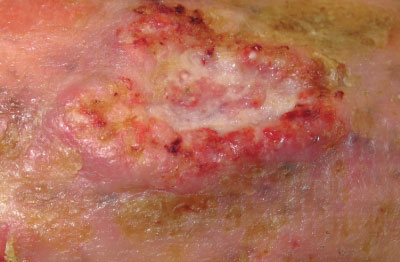
Cutaneous squamous cell carcinoma: Invasive keratinocytic skin cancer typically described as an enlarging scaly or crusted ‘volcano-like’ nodule or irregular infiltrated plaque. Squamous cell carcinomas grow over weeks to months and are often tender or painful and may ulcerate (Figure 3). Dermatoscopy generally shows a central scale/crust and peripheral white structures or structureless zones. A pink colour and polymorphous vessels are most often seen in fast-growing tumours.

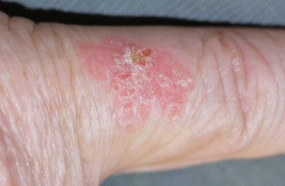
Cutaneous squamous cell carcinoma in situ: Also known as intraepidermal squamous cell carcinoma. A red or pigmented, slowly growing, scaly plaque; often multiple (Figure 4). Most common on sun exposed sites but may arise anywhere. Dermatoscopy shows clusters of rounded vessels (red dots, clods, coils); vessels and pigment are often in linear array.

Melanoma and excision terminology

Melanoma: The malignant proliferation of melanocytes usually within the epidermis (in situ) or dermis (invasive). Most often arises from normal skin but approximately one-third arise within a melanocytic naevus (precursor lesion). Dermatoscopic features are characterised by chaos and one or more clues to malignancy. Also see “Subtypes of melanoma”.

Narrow complete excisional biopsy: An excision biopsy with 2 mm margins, that encompasses the entire lesion and is of sufficient depth to avoid transection at the base.5

Wide local excision: Re-excision including an extra margin of tissue that is removed from the original excision site following a positive pathology report for melanoma. Margins vary depending on Breslow thickness (distance of the deepest point of the tumour from the granular layer of the epidermis).

For further information and for images of specific lesions, see: https://dermnetnz.org/
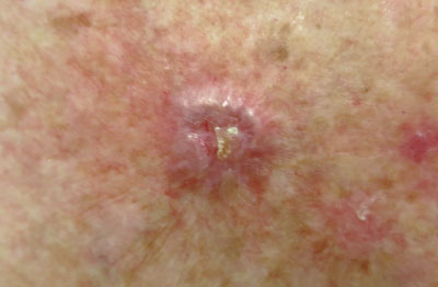
Figure 2:
Nodular basal cell carcinoma (image supplied by DermNet NZ).

Figure 3:
Poorly differentiated and ulcerated squamous cell carcinoma (image supplied by DermNet NZ).

Figure 4:
Cutaneous squamous cell carcinoma in situ (image supplied by DermNet NZ).

Acknowledgement

Thank you to the Te Aho o Te Kahu – Cancer Control Agency and their Melanoma Working Group, in particular Dr Susan Seifried (general surgeon), Dr Mark Foley (general practitioner), Dr Christopher Adams (plastic and reconstructive surgeon) and Dr Melissa James (radiation oncologist), and Dr Amanda Oakley, Adjunct Associate Professor and Consultant Dermatologist, Waikato DHB and for expert review of this article.

Article supported by Te Aho o Te Kahu – Cancer Control Agency.

N.B. Expert reviewers do not write the articles and are not responsible for the final content. bpacnz retains editorial oversight of all content.


Niall Holland 16 Dec 2021 15:47

Please login to make a comment.

Made with by the bpacnz team

Partner links