Key practice points:
- Impetigo, also known as “school sores”, is a common,
highly contagious bacterial infection of the skin
- Impetigo is usually diagnosed clinically. Swabs
may be required for recurrent infections, treatment failure with oral antibiotics, or where there is a community outbreak.
- First-line treatment of localised non-bullous impetigo
should focus on good skin hygiene measures and use of a topical antiseptic
- Use of a topical antibiotic is discouraged, but
it may be considered for a small area of localised infection if topical antiseptics have been trialled and were unsuccessful
or were not appropriate due to location of infection (e.g. around the eye)
- Oral antibiotics are recommended for people with
more extensive infection (i.e. more than three lesions/clusters), bullous impetigo, systemic symptoms or when topical
treatment is ineffective
Impetigo can affect people of any age, but it most commonly occurs in young children (i.e. aged two to six years).1 Staphylococcus
aureus and Streptococcus pyogenes, either alone or together, are the most common causes of impetigo.1 Impetigo
can occur in an area of previously healthy skin or at the site of minor trauma that disrupts the skin barrier, such as
a graze, scratch or eczema.2
Impetigo is highly contagious and can be transmitted by direct contact, often spreading rapidly through families, day-care
or schools.1
Impetigo is more common in:1 ,3–6
- Hot humid weather
- Conditions of poor hygiene, e.g. overcrowding, or close physical contact, e.g. contact sports
- People who have skin conditions or experience trauma that impairs the normal skin barrier, e.g. eczema, scabies, fungal
skin infections, abrasion, insect bites
- People with diabetes mellitus
- People who are immunocompromised
- People who use intravenous drugs
There are two types of impetigo: bullous and non-bullous
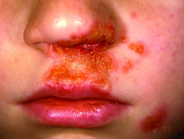
Non-bullous impetigo (Figure 1) is the most common variant, and is usually caused
by S. aureus but in some cases may be caused by S. pyogenes.1 Lesions begin as a vesicle that
ruptures and the contents dry to form a gold-coloured plaque on the skin.1 These lesions are often 1–2 cm in
diameter and most frequently affect the face (especially around the mouth and nose) and limbs.3 Systemic signs
are not usually present, however, with more extensive impetigo, fever and regional lymphadenopathy may occur.2, 7
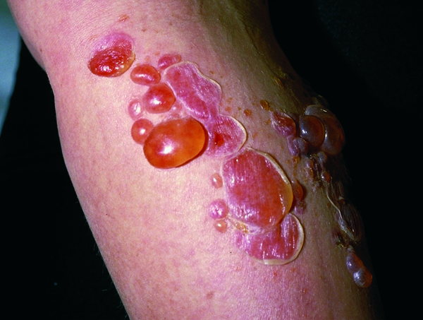
Bullous impetigo (Figure 2) is only caused by S. aureus and accounts
for approximately 10% of cases, most often seen in infants.1, 2 It is characterised by larger fluid-filled blisters
that rupture less easily than blisters from non-bullous impetigo, leaving a yellow-brown crust.1 Systemic signs
of infection such as fever and lymphadenopathy are more likely to occur and the trunk is more likely to be affected.2
N.B. Ecthyma is a deep tissue form of impetigo. It is characterised by crusted sores beneath which ulcers form with
a “punched out” appearance.8 It is more common in children, older people and immunocompromised people or
in conditions of poor hygiene and hot humid weather.8 Treatment follows the same guidelines as impetigo,
but oral antibiotics are usually required.9
 |
 |
| Figure 1. Non-bullous impetigo. Image provided by DermnetNZ |
Figure 2. Bullous impetigo. Image provided by DermnetNZ |
Impetigo is usually diagnosed clinically
Impetigo can be diagnosed on clinical examination and initial treatment decisions are rarely based on the results of skin
swabs.3 Swabs may be required for people with recurrent infections, treatment failure with oral antibiotics,
or where there is a community outbreak and the cause needs to be identified.5 For people with recurrent impetigo,
nasal swabs can identify staphylococcal nasal carriage requiring specific management.5
Key points:
- The aims of treatment are to clear the eruption and prevent the spread of the infection to others
- Good skin hygiene measures and a topical antiseptic are first-line for children with mild to moderate impetigo
- Due to increasing resistance, infectious disease experts recommend that topical antibiotics should have a very limited
role in clinical practice10, 11
- Oral antibiotics are suitable for people with more extensive or recurrent infection5
- Combination treatment with a topical and oral antibiotic should not be offered5
- Underlying conditions e.g. eczema need to be treated as well to reduce the risk of recurrent impetigo4
For further information, see:
Topical antiseptic is the initial treatment for localised patches of impetigo
A topical antiseptic, e.g. hydrogen peroxide 1% or povidone-iodine 10%, applied two to three times daily, is the first-line
treatment for localised, uncomplicated non-bullous impetigo (e.g. three or less lesions/clusters).4, 5 The crusts
on the lesions should be removed with warm water before any topical preparation is applied (see: “Advice
for patients with impetigo”).2 Remind parents/caregivers to wash their hands before and after application.
Five days of topical antiseptic treatment is usually sufficient for treating impetigo.5 This can be increased
to seven days depending on the severity and number of lesions.5
Advice for patients with impetigo or their caregivers4, 20
To remove crusted areas:
- Use a clean cloth soaked in warm water to gently remove crusts from lesions. Wash the cloth after use.
To prevent the spread of infection:
- Children should stay away from day-care or school until the lesions have crusted over or they have received at least
24 hours of antiseptic or antibiotic treatment*. This may not be necessary for older children (e.g. secondary
school) who are able to minimise risk of transmission by avoiding physical contact with others.
- Avoid close contact with other people, e.g. siblings and other family members, contact play with other children
- Use separate towels, face cloths, clothing and bathwater until the infection has cleared. Disinfect linen and clothing
by hot wash, hot dry or ironing.
- Follow the “clean, cut (nails) and cover” message, which also can apply to people with other skin infections or injuries:
- Use hand sanitiser and/or careful washing with household soap and water, several times daily
- Keep children’s fingernails cut short to prevent bacteria spreading from one part of the body to another through scratching
- Cover the affected areas with a breathable dressing and wash hands after touching patches of impetigo or applying topical treatments
*As days off school equate to increasing educational disparity and parental time off work (often
without pay), families should be educated and supported in strategies to prevent skin infections.
For further information, see:
https://www.kidshealth.org.nz/how-stop-skin-infections
Use of topical antibiotics is discouraged
Topical antibiotics are rarely indicated for use in skin infections due to bacterial resistance and the potential for
contact dermatitis.4 However, there may be some instances where a topical antibiotic is considered for treating
a small, localised area of impetigo, such as if a topical antiseptic is unsuitable (e.g. impetigo around the eyes) or has
been ineffective.5 Fusidic acid should be prescribed unless antibiotic sensitivities (if known) indicate that
resistance is present. Mupirocin (partly funded) is reserved for treating MRSA (see: “Impetigo caused by
MRSA”).10
Impetigo caused by MRSA
The prevalence of impetigo caused by methicillin-resistant S. aureus (MRSA) is unknown, but is likely to
be increasing.12 In August 2017*, 956 MRSA laboratory isolates were reported in New Zealand, equating
to a period-prevalence rate of 19.9 patients with MRSA per 100,000 population.13 This is double that of isolates
from 2009, but rates have remained relatively stable over the last four years.13 Some community strains of
MRSA show increasing resistance to fusidic acid, while resistance rates of mupirocin are decreasing concurrently with declining
dispensing rates.11,14 Oral trimethoprim + sulfamethoxazole, tetracyclines or clindamycin are usually effective
against MRSA.15
* 2017 is the latest data as this survey has not been conducted since.
Oral antibiotics should be used for multiple lesions or if topical treatment is ineffective
Oral antibiotics are recommended to treat patients with more than three to five lesions/clusters, bullous impetigo, systemic
symptoms or when topical treatment is ineffective.5 Flucloxacillin is the first-line choice as it is effective
against S. aureus and group A streptococci.4, 5
Trimethoprim + sulfamethoxazole or erythromycin can be used if MRSA is present or the patient is allergic or intolerant
to flucloxacillin. Cefalexin is another option if flucloxacillin is not tolerated.4, 5
A five day course of oral antibiotics is generally sufficient, but can be increased to seven days depending on the severity
and number of lesions.5 If treatment is unsuccessful after this time, medicine adherence should be checked and
swabs can be taken to detect sensitivities.5
Refer to “Antibiotics: choices for common infections” for dose and regimen information. Available from:
https://bpac.org.nz/antibiotics/guide.aspx
Prevention of recurrent impetigo infections
Recurrent infection may result from the nasal carriage of causative microorganisms, close contact with others or from
fomite colonisation e.g. bed sheets, towels and clothing that may be shared.4, 16 If nasal carriage is suspected,
a nasal swab is required to confirm this and to establish antibiotic susceptibility. A topical antibiotic should be applied
inside each nostril, three times daily for seven days. The choice of antibiotic will be guided by sensitivities (from swab
result). All household members and close contacts should also be treated.4
For further information on decolonisation, see:
https://bpac.org.nz/2017/topical-antibiotics-2.aspx
Post-streptococcal glomerulonephritis, which can lead to renal failure, is a rare complication of streptococcal
impetigo.4 Treatment of impetigo may not prevent susceptible people developing this complication.15 Prevalence
of post-streptococcal glomerulonephritis is highest in primary school aged children, particularly males and people of Māori
and Pacific descent.17
Scarring may occur in people with more severe impetigo, when lesions extend deeper into the dermis.8 N.B.
In milder cases of impetigo, healed lesions may result in changes in skin pigmentation, however, this should resolve over
time.2, 3
Soft tissue infection such as cellulitis may occur.4
Staphylococcal scalded skin syndrome is characterised by red blistering skin which leaves an area that
looks like a burn once the lesions have ruptured. Children aged under five years, particularly neonates, immunocompromised
people or those with renal failure are most at risk of this complication.18
Streptococcal toxic shock syndrome is a rare complication of impetigo. It is more commonly seen in healthy
people aged 20 to 50 years, despite children, immunocompromised and elderly people being more susceptible to impetigo. Symptoms
include fever, rash, hypotension and erythematous rash.19
Rheumatic fever is rarely linked to skin infections however it can occur when group A streptococci found
on the skin moves to the throat.4


