Published: 25th September, 2026
Key practice points
- Consider hip dysplasia in any young patient with insidious, activity-related groin pain, particularly females and especially if they report mechanical symptoms (clicking, catching, a sense the hip is “giving way”). These features should raise suspicion beyond a straightforward groin strain.
- A normal newborn hip check or infant hip ultrasound result does not exclude dysplasia later in life. A considerable proportion of adolescents and young adults with symptomatic dysplasia have had entirely normal infant screening.
- Features from the patient’s history can be combined with three targeted examination tests (the anterior apprehension test, FADIR and FABER) to build clinical suspicion of hip dysplasia, before requesting imaging
- If X-ray is indicated, request a supine AP pelvis and either a false-profile (preferred) or frog-leg lateral view. Look for a lateral centre-edge angle < 20 – 25° and/or a Tönnis angle > 10° on the report.
- If dysplasia is confirmed and the patient is symptomatic, refer them promptly for orthopaedic assessment as surgical joint-preservation options narrow considerably once secondary osteoarthritis develops
- While awaiting specialist assessment, the patient can be prescribed NSAIDs for pain, advised to modify activities and referred to physiotherapy for exercises targeting deep hip and core stabilisers; this is short-term management and insufficient alone unless the patient has only mild dysplasia
- Periacetabular osteotomy (PAO) preserves the natural joint and works best before the onset of arthritis; total hip arthroplasty (THA) is also an option, but subsequent revision is highly likely to be required in younger patients
- Untreated symptomatic hip dysplasia significantly increases the risk of early osteoarthritis. Timely diagnosis and management can change a patient's lifetime trajectory of disease and disability.
Hip and groin pain in adolescents and young adults is common, and in almost all cases the diagnosis is a muscular strain, hip flexor tendinopathy or benign activity-related pain. However, in a small group of these patients, there is an underlying structural cause – hip dysplasia. The presentation can closely resemble these more common conditions, making it easy to overlook on a first assessment. Population-based studies suggest that hip dysplasia is present more often than it is documented in the radiology report: a Scandinavian cross-sectional study identified hip dysplasia in 5.2% of 1,870 adult radiographs, despite it only being recorded in the radiology report for approximately 7% of these patients (when the relevant angles were specifically calculated).1 Consistent with these findings, the average time from symptom onset to a confirmed diagnosis of acetabular (hip) dysplasia has been reported at more than five years.2
Part of what makes the diagnosis of hip dysplasia so challenging is that many young people present with symptoms that overlap with far more common causes of groin pain. The classic picture is a skeletally mature, young, active patient, often but not exclusively female, with insidious, activity-related groin and/or lateral hip pain.3 There is usually no specific injury that the pain can be attributed to, no features that immediately stand out, and a physical examination that, without specific hip-focused testing, looks reassuringly normal. Recognising specific diagnostic features (see: “What is hip dysplasia?”), can help identify a small number of patients with potential hip dysplasia, from the much larger group who likely have a straightforward soft-tissue strain.
So, why does this distinction matter so much for young patients? The reason is that the surgical treatments that best preserve a young person's natural hip joint work far better before secondary osteoarthritis develops, so earlier recognition has a real, measurable effect on long-term joint survival.4
What is hip dysplasia? The underlying mechanical process
A normally formed acetabulum is a deep, well-rounded socket that covers most of the femoral head, distributing the forces of standing, walking and running across a wide surface of articular cartilage. In a person with hip dysplasia, the acetabulum is shallow and often steeply angled, most often resulting in under-coverage of the femoral head laterally, anteriorly or both.
This has two connected mechanical consequences:5, 6
- A smaller weight-bearing surface concentrates load. With less bony roof available to share the work, the same body weight and muscular forces are transmitted through a smaller area of cartilage and rim, increasing contact stress at the acetabular edge, the weakest point.
- A shallow socket is an unstable socket. Without a deep, well-contained cup for the femoral head, it is freer to migrate within the joint during movement, particularly toward the under-covered region. The acetabular labrum, which in a normally covered hip has only a minor stabilising and sealing role (to prevent fluid loss from the joint space), is recruited to compensate for this instability, taking on a mechanical, load-bearing job it was not designed for, which over time results in hypertrophy.
This compensatory overload is typically when hip dysplasia starts to cause pain. Chronic excess loading at the chondrolabral junction can result in labral tearing, fraying and eventual detachment, sometimes described as an “acetabular rim syndrome”. A torn labrum loses its seal, i.e. the mechanism by which the joint normally maintains a thin, low-friction layer of synovial fluid between cartilage surfaces. This accelerates cartilage wear, prompting the release of inflammatory breakdown products that further degrade joint lubrication.5, 6 This process creates a pathological cycle that leads to secondary osteoarthritis, with onset occurring often decades earlier than would be expected with typical age-related wear.6 Hip dysplasia is the main cause of hip osteoarthritis in patients aged under 60 years.6
Femoral version also contributes to joint stability and is often overlooked. In addition to acetabular shape, the rotational alignment of the femur itself, known as femoral version, contributes to how well the femoral head is contained. Increased femoral anteversion, i.e. > 20° (structurally normal is 10 –15°) can co-exist with acetabular dysplasia. One study of patients aged 13 – 49 years with symptomatic acetabular dysplasia who underwent pelvic CT scans during surgical planning, found that increased femoral anteversion was also present in approximately one-third of the patients.7 An excessively anteverted femur effectively reduces how much of the femoral head is covered by the acetabulum during extension and external rotation, exacerbating anterior instability even when acetabular coverage looks only mildly abnormal on plain X-rays. This is one reason why two patients with a similar lateral centre-edge angle can present with different degrees of clinical instability.
It's important to recognise that hip dysplasia is a problem of instability as much as shape. Many patients, particularly young, athletic females in whom this condition is over-represented, also have generalised ligamentous laxity (i.e. they are very flexible). This soft-tissue hypermobility compounds the structural under-coverage and helps explain why a hip can look only mildly shallow on X-ray yet still feel profoundly unstable to the patient.
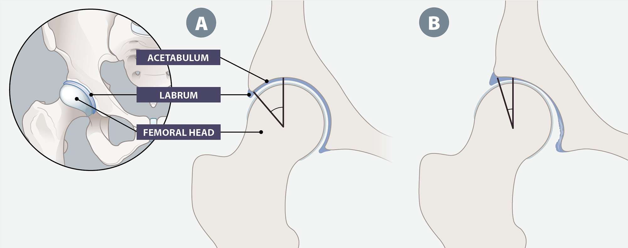
A: “Normal hip”: deep, rounded acetabulum cupping the femoral head; centre-edge angle near 35°; load spread evenly; a thin, unremarkable labrum.
B: “Dysplastic hip”: shallow, steep acetabulum, femoral head displaced and only partly covered; centre-edge angle under 20°; load concentrated on the lateral rim; the labrum hypertrophied.
Adolescent-onset hip dysplasia: A different road to the same destination
It is often assumed that hip dysplasia in an adolescent or young adult must simply be a case missed by infant screening. However, this is not necessarily the case, and the reality is more nuanced. Understanding this provides important context for how primary care clinicians should think about risk in these patients.
There are two recognised pathways to symptomatic hip dysplasia in adolescents and young adults:6
- Residual dysplasia from developmental dysplasia of the hip (DDH). The hip was recognised as unstable or dysplastic in infancy, and may have been treated with a harness, splinting or surgery, but did not fully remodel to a normal shape. Even after apparently successful treatment of infant DDH, a small proportion of adolescents and young adults have hips that remain mildly dysplastic at skeletal maturity. It is also possible that the dysplasia was not identified during screening in infancy, even though it was present.
- Adolescent (or late)-onset dysplasia. The hip was stable and structurally normal at birth (confirmed with clinical examination and, where performed, a normal ultrasound), but a shallow, under-covering acetabulum becomes apparent only later, as the child grows, begins walking and progressively loads the joint through childhood and adolescence.
Adolescent-onset of hip dysplasia is increasingly recognised as a distinct developmental process to infant DDH.6 The problem is that it is extremely difficult to predict. In one German cohort of adult patients with hip dysplasia who underwent a periacetabular osteotomy (see: “Surgical treatment options for hip dysplasia”), approximately half of the 94 participants who were able to provide conclusive infant screening results had a normal ultrasound recorded.8 Germany has a universal neonatal ultrasound screening programme. In an American study, approximately 85% of the 68 patients aged 13 – 47 years who presented with symptomatic dysplasia would not have met the current criteria for risk-factor-based selective ultrasound screening as infants, if applied retrospectively (this screening approach is also used in New Zealand, Australia and the United Kingdom).9 Even with universal clinical examination at birth and again at six to eight weeks, cohort studies continue to report infants who go on to have a late diagnosis of hip dislocation, with a mean delay to diagnosis of well over one year.10
The classic infant risk factor for DDH is breech presentation. However, this is less common in people with late-onset hip dysplasia than other factors, e.g. female sex, firstborn status and a family history of hip osteoarthritis (dysplasia may have been an unrecognised underlying cause).9 None of these factors are a reason for closer neonatal screening, which helps explain why the condition is so often missed.
It is estimated that between one- and two-fifths of cases of adult hip osteoarthritis are related to unrecognised or under-treated developmental hip dysplasia.11 So, rather than being a rare occurrence, hip dysplasia is a meaningful, under-appreciated contributor to osteoarthritis, a condition that primary care clinicians manage every day.
Key practice point: A normal newborn hip check or ultrasound does not guarantee that the hip will develop normally, and it does not exclude dysplasia as a cause of hip pain many years later.
Suspicion of hip dysplasia should be increased in a young, active, otherwise well patient presenting with slow-onset, activity-related groin and/or lateral hip pain.6 This can occur in any adolescent or young adult, although the classic demographic is a female involved in dance, gymnastics, football or another pivoting, impact or deep-flexion sport.2 Unlike with a muscular strain, there is usually no single traumatic event associated with pain onset; typically, some pain has always been present and worsened gradually, sometimes after an unremarkable increase in training load or another activity, e.g. an active holiday, taking up running or weight-bearing exercise involving lunging or squatting.12
Ask about:2, 3, 12
- Site and quality of the pain – deep, anterior groin pain is most common, but lateral hip and buttock pain can also occur. As “hip pain” is often interpreted broadly, ask the patient to localise their pain with a pointed finger rather than an open hand.
- Look for the “C-sign” – many patients cup the lateral hip between their thumb and fingers in the shape of a “C” when describing their pain. This is a non-specific but recognisable feature of intra-articular hip pathology that should prompt further assessment.
- Mechanical symptoms – clicking, catching, locking or a sensation of the hip “giving way” suggest labral pathology, which is much more likely to occur secondary to dysplasia than as an isolated finding
- Aggravating positions – pain is often worse with prolonged standing or walking (i.e. weight-bearing), prolonged sitting, pivoting or cutting movements, deep flexion (e.g. putting on shoes and socks, sitting on a low chair, squatting) and end-range extension with external rotation (e.g. a dance arabesque, a goalkeeper's dive, hurdling)
- Instability sensations – the patient may describe a feeling of apprehension that the hip might subluxate, particularly when their leg is extended and rotated outward
- Infant/childhood hip history – ask about neonatal hip screening, use of a harness or brace in infancy, relevant symptoms (e.g. a “clicky hip”, problems sitting cross-legged), although the absence of any such history does not exclude the diagnosis
 Red flags for acute management
In most cases, hip pain in an adolescent or young adult is not an emergency. However, there are some clinical scenarios that require more acute management, including:
- Any patient who has fever, an acutely hot or swollen hip or refusal to weight bear; consider septic arthritis or osteomyelitis and arrange acute referral to secondary care
- An early to mid-adolescent patient with an antalgic or externally-rotated gait and groin, hip or knee pain, especially if overweight; consider a slipped upper femoral epiphysis. A positive Drehmann sign (i.e. obligatory external rotation on passive hip flexion) warrants same-day orthopaedic referral, due to the high risk of further epiphyseal slip.13
- A patient who is an endurance athlete with point tenderness over the femoral neck or groin and a history of disproportionate night pain, particularly if accompanied by menstrual disturbance, disordered eating or low energy availability in females; consider a femoral neck stress fracture and screen for Relative Energy Deficiency in Sport (REDS)14
For further information about REDs, see: bjsm.bmj.com/content/57/17/1073.long
- Any patient with unexplained weight loss, night sweats or a personal history of malignancy; consider a bony or soft tissue tumour
Hip examination: Three tests worth learning
A hip examination in primary care should be carried out systematically. It does not require any special equipment.
Start by assessing the patient’s gait and single-leg stance: a positive Trendelenburg sign (i.e. pelvic drop on the unsupported side) reflects abductor dysfunction, which is common in people with chronic dysplasia because of the lateralised, shortened abductor lever arm.15
Next, work through the three provocative tests as follows:14
1. Anterior apprehension test (hyperextension–external rotation): assesses for micro-instability
- Position the patient supine at the end of the examination table
- Ask them to flex their unaffected hip and hold knee to their chest (flattens lumbar lordosis and stabilises the pelvis)
- Allow the affected leg to drop off the edge of the table into extension
- Gently externally rotate the extended hip
Interpretation: a positive test reproduces anterior groin pain and/or apprehension, instability or visible guarding as the patient contracts to protect the hip, reflecting anterior capsular and labral laxity from under-coverage.16 The reported sensitivity of the anterior apprehension test is 71%, with a specificity of approximately 85%.16
Other provocative manoeuvres for assessing micro-instability include the abduction-extension-external rotation and prone external rotation tests.15 A positive result for all three tests correlates strongly with instability diagnosed with arthroscopy; a single clearly positive test in the right clinical context is enough to justify imaging.16
2. FADIR test (flexion, adduction, internal rotation, also referred to as FADDIR) – assesses for femoroacetabular impingement (FAI) and labral pathology15
- With the patient supine, passively flex the hip and knee to 90°
- Adduct the hip across the midline
- Internally rotate the hip
Interpretation: reproduction of anterior groin pain is a positive test.15 FADIR tested at 90° of hip flexion is sensitive for detecting FAI (approximately 60 – 100%) but lacks specificity, so is better used to build suspicion, or to reassure if negative, than to distinguish dysplasia from FAI or an isolated labral tear.15
3. FABER test (flexion, abduction, external rotation, also called Patrick's test or sign) – assesses for FAI and labral pathology, as well as sacroiliac joint pathology15
- With the patient supine, rest the ankle of the test leg on the contralateral knee, forming a “figure-4” position
- Stabilise the contralateral anterior superior iliac spine with one hand
- With the other hand, apply gentle, steady downward pressure on the medial aspect of the flexed knee towards the examination table
Interpretation: decreased range of motion relative to the contralateral hip or reproduction of pain is a positive result.15 Groin pain suggests intra-articular hip pathology (e.g. labral tear, dysplasia, FAI), while pain localised posteriorly on the contralateral side suggests sacroiliac joint dysfunction.15 Comparing knee-to-table distance side to side can be broadly interpreted: dysplastic, hypermobile hips often descend further than expected, whereas hips with FAI are frequently restricted. As hip rotation range can also be influenced by femoral version (natural rotational angle of the femur) in addition to acetabular shape, an unusually large or restricted arc of rotation is a general sign of combined pathology rather than a way of separating the two possibilities.15
Requesting initial imaging: Ordering and interpreting X-rays
Plain radiography is the first-line investigation if dysplasia is clinically suspected. Request a supine AP pelvis and a false-profile (Lequesne) view. If a false-profile view is not readily available, request a frog-leg lateral view and clearly indicate suspected dysplasia on the request form, so the radiologist comments specifically on anterior coverage. Requesting optimal views from the outset helps to avoid one of the most common reasons hip dysplasia diagnosis is delayed.1
Plain films cannot reliably quantify femoral anteversion; advanced imaging, e.g. a MRI or MR arthrogram for labral assessment or a CT for femoral version, is usually requested in secondary care once surgical planning begins.
Local referral protocols and criteria may influence this process; check HealthPathways. In some areas it may be necessary to discuss with a paediatric or orthopaedic specialist before requesting certain types of imaging. For information about relevant X-ray referral criteria, see: radiology.carepathways.tewhatuora.govt.nz/national/community-referred-radiology
Read more about X-ray
The baseline X-ray for an adolescent or young person with symptomatic suspected hip dysplasia is a supine anteroposterior (AP) view of the whole pelvis correctly centred (i.e. coccyx aligned over the pubic symphysis, obturator foramina symmetrical).17, 18 A standing, weight-bearing view is recommended in some literature on the basis that it captures the hip under physiological load.17 In practice, however, standing pelvis radiographs are technically demanding to perform consistently, and variability in radiographer positioning can undermine the reliability of the measurements. A carefully centred supine view is more consistently achievable in routine practice; a dedicated reliability study reported equivalent lateral centre-edge angle measurements between supine and standing positions.19
Anterior acetabular deficiency is common in adolescents and young people with dysplasia and can co-exist with an apparently preserved lateral centre-edge angle.17, 18 As the AP view alone can only be used to quantify lateral coverage, a second orthogonal view is also required, i.e. either:
- False-profile (Lequesne) view – the patient stands with their affected hip against the cassette, and their pelvis rotated 65° from the coronal plane.17 This directly measures the anterior centre-edge angle (angle of Lequesne) and is the preferred second view when dysplasia is the most likely diagnosis.18
- Frog-leg lateral view – the patient is supine, with their hips flexed to approximately 40 – 45°, abducted and externally rotated.18 This view is simpler and more widely available and is used to assess femoral head sphericity and head–neck offset (useful for co-existing cam morphology, i.e. a bony bump or flattening on the femoral head) but does not reliably quantify anterior coverage.17, 18
Interpreting radiology results
Key features to look for in the patient’s radiology report include:
- Lateral centre-edge angle (LCEA) of Wiberg: ≥ 25° is normal; 20 - 25° is generally classed as “borderline dysplasia”, with < 20° considered diagnostic for dysplasia20, 21
- Tönnis (acetabular roof) angle: 0 – 10° is normal; > 10° indicates dysplasia20
- Anterior centre-edge angle (angle of Lequesne): < 20° indicates anterior under-coverage even if the lateral centre-edge angle is preserved, a pattern that frequently explains a reassuring single-view AP film17
- Mentions of a break in Shenton's line*, a flattened or incongruent sourcil (acetabular roof), and early secondary osteoarthritic change(s) (e.g. joint space narrowing, subchondral sclerosis).17 The presence of any of these features supports referral, regardless of the reported angle values.
If the report is purely descriptive, e.g. “no acute bony abnormality”, and does not quote these angles, it is recommended to request that they are added (they are not always routinely calculated). Population-based data suggest that dysplasia is more common than standard radiographic reporting suggests; in one New Zealand study of 68 patients who underwent periacetabular osteotomy for symptomatic hip dysplasia (see: “Surgical treatment options for hip dysplasia”), the diagnosis was documented in the initial radiology report for only approximately 50% of patients.22 This trend has also been reported overseas.1
* The imaginary line that follows the arc formed by the superior border of the obturator foramen and the medial margin of the femoral neck; a disruption to the line can indicate femoral displacement17
Summing up the case: Distinguishing hip dysplasia from common mimics
Table 1. Key clinical and diagnostic features of hip dysplasia and other common hip pathologies.
|
| Hip dysplasia |
FAI syndrome (femoroacetabular impingement)23 |
Adductor-related groin pain24 |
Inguinal-related groin pain (i.e. a “sports hernia”)24 |
Typical patient characteristics |
Young, active person (usually female), often with generalised ligamentous laxity (may report being “very flexible”), e.g. dancers, gymnasts, footballers |
Young, active person involved in pivoting and/or deep-flexion sports; more frequently affects males |
Person involved in kicking and/or change-of-direction sports, e.g. football, hockey |
Person involved in sprinting and/or twisting sports, e.g. football, rugby, ice hockey; predominantly affects males |
Onset |
Insidious, atraumatic; may have a history of infant developmental dysplasia of the hip |
Insidious, atraumatic |
Often acute, e.g. during a specific kick or twist |
Insidious, worsening over weeks to months |
Pain pattern |
Deep anterior groin, sometimes extending to the lateral hip/buttock; “C-sign” |
Deep anterior groin; “C-sign” |
Localised to the proximal adductor origin/medial thigh |
Deep groin/lower abdominal wall; may radiate toward the perineum |
Mechanical symptoms |
Common, e.g. clicking, catching, “giving way” |
Common, e.g. clicking, catching, locking |
Absent |
Absent |
Aggravating factors |
Prolonged standing, walking or sitting, pivoting, deep flexion |
Deep flexion, prolonged sitting, pivoting |
Resisted adduction, kicking, sprinting |
Sit-ups, coughing/sneezing, sprinting, twisting |
Key examination findings |
Positive anterior apprehension test often with increased hip range of motion; Trendelenburg sign may be present |
Positive FADIR test with reduced internal rotation in flexion |
Adductor tenderness with pain on resisted adduction |
Tenderness over the conjoint tendon/pubic tubercle and pain on resisted sit-up with normal hip examination |
Radiological findings |
Lateral centre-edge angle (LCEA) of < 20 – 25° and/or Tönnis angle > 10° |
Cam and/or pincer morphology on lateral views |
Usually normal, if performed; in most cases, diagnosis is clinical |
Usually normal; imaging mainly excludes hip-related causes |

In most cases, adolescent or young adult patients with radiologically confirmed hip dysplasia require secondary care referral; the next steps depend on their clinical circumstances and local referral criteria and resources. Surgery is often required - the exception may be patients with mild dysplasia. Primary care clinicians can support patients by explaining the treatment options and providing conservative management while they wait for specialist assessment. The primary care team also has an ongoing role in post-surgical care and general lifestyle management.
Indications for secondary care referral
Refer any adolescent or young adult with symptomatic, radiologically confirmed dysplasia (i.e. lateral centre-edge angle < 20 – 25° and/or Tönnis angle > 10°) for orthopaedic assessment; ideally for the attention of an orthopaedic surgeon with an interest in hip preservation (or directly if private referral).
 If any of the following factors are present, detail them in the referral, with the aim of achieving a fast-track review:
- Confirmed dysplasia with ongoing pain or functional limitation despite an adequate trial of conservative care (see below)
- Instability signs on examination (e.g. a positive anterior apprehension test) alongside confirmatory imaging
- Mechanical symptoms suggesting a labral tear (e.g. clicking, catching, locking, or a sense of the hip giving way)
- Any secondary osteoarthritic change already visible on X-ray (e.g. joint space narrowing, subchondral sclerosis): joint-preserving surgery may no longer be suitable. Prompt assessment is required both to confirm what treatment options remain and because further delay will worsen the joint space narrowing.
- A young, physically active patient whose goals include returning to pivoting or impact sport (e.g. an athlete or dancer)
- Bilateral dysplasia or a strong family history, which may warrant broader assessment
Follow your usual local referral process for orthopaedic assessment. Where the radiology report includes angle measurements, quote these in your referral letter, along with a clear description of your clinical suspicion of instability or dysplasia, to support accurate triage.
Management while awaiting referral: Conservative care
A trial of conservative care is appropriate for most patients while they wait for specialist assessment. Conservative care does not correct the underlying structural deficiency associated with hip dysplasia, but it can meaningfully reduce symptom burden and joint load.25 For patients with milder, borderline dysplasia, it may be the only treatment needed.
Physiotherapy. Refer for physiotherapy specifically targeting deep hip and pelvic stabilisers; generic gym-style strengthening is not sufficient. Expert consensus on non-operative rehabilitation for hip dysplasia recommends progressing through local muscle control (e.g. transverse abdominis activation, gluteal bridging, side-lying hip abduction against resistance), then global control (e.g. single-leg stance, single-leg squat, lateral step-downs) and low-demand lumbopelvic control work (e.g. supine marching, the bird-dog exercise).25 Early isolated hip flexor strengthening is generally avoided as the iliopsoas is often already overloaded from compensating for anterior instability; it is generally only considered following improvements in posterolateral hip strength and hip joint irritation.25
Activity modification. Advise patients to reduce, but not necessarily stop, repetitive deep flexion, pivoting and high-impact loading while they have active symptoms, e.g. modifying training volume in dancers, gymnasts and footballers. Activities that do not increase pain can be continued, however, those that do should be modified, e.g. reducing intensity or frequency, or temporarily stopping (with reinitiation when tolerance improves).25 Complete cessation of sport is rarely necessary or sustainable.
NSAIDs. Short-term, judicious use of a NSAID can settle symptomatic flares. e.g. 2 – 4 weeks at the lowest effective dose, reviewed for effect. However, long-term reliance on NSAIDs should be avoided, due to the risk of adverse effects and because it can mask progressive mechanical symptoms that should instead prompt earlier specialist review. Be particularly cautious with use of NSAIDs in athletes with disordered eating or low energy availability, who are at increased risk of gastrointestinal and renal adverse effects.
Surgical treatment options for hip dysplasia
There are two main procedures for the surgical management of hip dysplasia in adolescents and young adults. Familiarity with these procedures is useful for primary care clinicians, so they can be explained to patients and their family/whānau.
Periacetabular osteotomy (PAO)
The Bernese (Ganz) periacetabular osteotomy (PAO) involves reorientating the acetabulum around an intact posterior column, improving femoral head coverage while preserving the natural joint.26 It is the preferred treatment option for patients with symptomatic dysplasia and little or no existing arthritis. Historically, patients aged over 40 years would have been excluded from this surgery due to inferior outcomes. However, more recent evidence suggests that outcomes now appear more closely related to the absence of pre-existing arthritis than to age itself.4 A PAO is a technically demanding, open operation, requiring a hospital stay, followed by six to eight weeks of protected, crutch-assisted weight-bearing, and approximately three to six months of structured rehabilitation before a return to unrestricted activity. Reported cumulative hip survivorship (i.e. not requiring conversion to a total hip replacement) in the original Bernese cohort was reported at approximately 88% at 10 years and 61% at 20 years post-procedure.27 At 30 years post-procedure, the absence of arthritis progression and pain was reported in approximately one-third of patients.27 The procedure also improves hip function and patient quality of life.4
It is important to note that isolated hip arthroscopy to trim a torn labrum, without addressing the underlying bony under-coverage, is not a substitute for PAO in patients with true structural dysplasia and can accelerate instability. In a small number of patients with significant co-existing femoral anteversion identified on CT, the specialist may combine PAO with a femoral derotation osteotomy to fully address the instability, however this possibility does not need to be covered in detail before referral.7
Total hip arthroplasty (THA)
A total hip arthroplasty (THA) is reserved for patients with established secondary osteoarthritis and those for whom joint preservation is no longer realistic. Dysplastic anatomy, including a shallow, often anteverted acetabulum and sometimes leg length discrepancy, makes THA in these patients technically more demanding than in those with typical age-related osteoarthritis, occasionally requiring specialised implants, bone grafting or a femoral shortening osteotomy. Outcomes in young patients with dysplasia are good, but implant survival is lower than in older populations as a result of the longer functional demand placed on the joint; around 87% at 10 years and 61% at 20 years in one long-term cohort of patients aged 35 years or younger at the time of procedure.28 Young adults undergoing a total hip arthroplasty should be counselled that it is highly likely they will need at least one revision during their lifetime.
The ongoing role of primary care
The role of primary care in the management of patients with hip dysplasia does not end with referral to a hip preservation specialist. Depending on the pathway a patient takes, primary care clinicians will continue to have an ongoing role in patient care.
While awaiting specialist review or once non-operative management is chosen. Support the patient with conservative care, continue to review symptom trajectory and reinforce their activity-modification plan and physiotherapy recommendations. Re-escalate the patient’s referral if their pain or mechanical symptoms worsen. Some patients with mild, borderline dysplasia may be successfully manged with conservative care without ever needing surgery, provided that the threshold for re-referral is clearly understood.
After periacetabular osteotomy. The surgical team is mostly responsible for post-operative care, however, patients will often attend general practice for wound concerns, breakthrough pain and questions, e.g. about extended thromboprophylaxis and crutch-assisted weight-bearing. Be alert to red flags suggesting infection, deep vein thrombosis/pulmonary embolism, or new neurological symptoms in the operated limb. Encourage adherence to the three-to-six month rehabilitation programme, emphasising the importance of not returning too early to high-impact activity.
After total hip arthroplasty. As for any patient undergoing a hip replacement, primary care shares responsibility with the surgical team for routine post-operative surveillance, venous thromboembolism prophylaxis review, and in the longer term, monitoring for signs of prosthetic joint infection or loosening. The threshold for imaging for suspected prosthetic joint loosening should be lower, given the relatively high revision rate for THA in this population.
Provide lifestyle advice to every patient
Regardless of the management pathway, two key points to discuss with every patient are:
- It is important to maintain a healthy body weight and avoid prolonged high-impact loading after surgery. Although PAO improves coverage, it does not fully restore the joint to “normal”, and a proportion of patients still eventually require a THA.
- Dysplasia has a recognised familial component. If they go on to have children, their history should be shared with their lead maternity carer (LMC) and the child’s paediatrician and primary care clinician. Patients can be reassured that their infants will be routinely screened and a positive family history of developmental hip dysplasia is a criterion for selective ultrasound screening in infants.29
Left untreated, symptomatic hip dysplasia will progress. The population-based Rotterdam Study reported a 4.3-fold increased risk of incident radiographic osteoarthritis in people with a centre-edge angle of < 25° compared to those with structurally normal hips.30 Studies of patients who underwent an early unilateral THA have shown that dysplastic hips develop degenerative change earlier and faster than either hips with FAI or contralateral structurally normal hips, particularly once even mild secondary degenerative changes become apparent on imaging.31
This is why early recognition of hip dysplasia is so important. Every year of diagnostic delay is a year in which the joint moves closer to the threshold where PAO is no longer appropriate, leaving THA, with the associated higher lifetime revision burden in young, active patients, as the only remaining surgical option. Patients identified with hip dysplasia and referred while the joint is still largely preserved have a different trajectory: i.e. pain relief, restored function, and, for many, decades of natural joint survival before THA is ever needed.26, 27 In the interim, maintaining a healthy body weight and avoiding prolonged high-impact loading are the two modifiable factors most within a patient's control while they wait for specialist assessment.
Resources for clinicians and patients
For clinicians:
- The New Zealand Orthopaedic Association Find a Surgeon directory covers both public and private practice and is searchable by location and subspecialty interest
- Ideally select an orthopaedic surgeon specialising in hips, along with paediatrics or sports. Periacetabular osteotomy is currently performed by a relatively small number of surgeons in New Zealand.
- A range of professional resources are available from The International Hip Dysplasia Institute website, including position statements on imaging and surgical indications. There is also a dedicated section with resources for paediatric and primary care clinicians.
For patients:
- The International Hip Dysplasia Institute website also has a range of resources for patients and their family/whānau, including specific information about adult hip dysplasia
- Miles4Hips is a patient-authored educational resource that aims to raise awareness and provide information about hip dysplasia, developed in partnership with the International Hip Dysplasia Institute