Published: 14th August, 2026
Key practice points
- COPD is estimated to affect more than 60,000 adults in New Zealand; Māori and Pacific peoples experience a higher burden of disease and are more likely to be admitted to hospital due to COPD
- The New Zealand COPD Guidelines were reviewed and updated in 2025, incorporating local consensus and expertise with both national and international evidence
- A formal diagnosis of COPD is based upon spirometry results in a patient with relevant symptoms and signs. A comprehensive patient history, physical examination and laboratory investigations, e.g. full blood count, BNP, support clinical suspicion and help to rule out other potential causes.
- A complete assessment and evaluation may require several consultations in primary care
- Non-pharmacological interventions continue to underpin COPD management, including smoking cessation, regular exercise, pulmonary rehabilitation and appropriate immunisations, e.g. COVID-19, influenza and pneumococcal vaccinations
- Pharmacological management provides symptom control and reduces the risk of exacerbations. It should be initiated in a stepwise approach depending on the patient’s clinical condition and presence of exacerbations.
- Most patients require a short-acting bronchodilator for relief of acute dyspnoea. This can be prescribed alone or alongside a long-acting bronchodilator depending on the patient’s frequency of symptoms.
- Funded options include a short-acting beta2-agonist (SABA), e.g. salbutamol or terbutaline, a short-acting muscarinic antagonist (SAMA), e.g. ipratropium, or a combination SABA + SAMA, e.g. salbutamol + ipratropium (preferred when a short-acting bronchodilator is prescribed alone)
- Prescribe a long-acting bronchodilator for any patient who experiences more than occasional dyspnoea. A long-acting muscarinic antagonist (LAMA), e.g. tiotropium, glycopyrronium or umeclidinium, is recommended for patients with persistent symptoms. N.B. A LAMA and a SAMA should not be used concurrently.
- Early escalation to combination treatment with a LAMA and a LABA is recommended for patients with persistent symptoms or who continue to experience exacerbations despite optimal LAMA monotherapy.
- Single inhaler combinations are preferred to optimise adherence; Special Authority approval for a LABA + LAMA single inhaler combination requires patients to have trialled a LAMA first
- Triple treatment (i.e. LAMA + LABA + an inhaled corticosteroid [ICS] or a single ICS + LAMA + LABA inhaler [preferred]) is recommended for patients with frequent or severe exacerbations
- To access a funded triple inhaler, patients must be using a LAMA + LABA (or ICS + LABA) inhaler and have any of the following:
- CAT score > 10
- Two or more exacerbations in the previous 12 months
- One exacerbation requiring hospitalisation in the previous 12 months
- Eosinophil count ≥ 0.3 × 10⁹ cells/L in the previous 12 months
- A blood eosinophil count ≥ 0.3 × 10⁹ cells/L suggests that the patient will likely benefit from ICS treatment
- An ICS + LABA inhaler (alone) is not recommended for COPD management unless the patient has a concurrent diagnosis of asthma
What’s changed?
This is a revision of a previously published article series: The optimal management of patients with COPD – Part 1: The diagnosis and Part 2: Stepwise escalation of treatment (BPJ 66, February, 2015), and standalone article, An update on the pharmacological management of stable COPD (June, 2020).
What’s new for this update:
- Combining information into one article for convenience
- Incorporation of latest recommendations from the New Zealand COPD Guidelines: 2025 Update
- Minor points from the Global Initiative for Chronic Obstructive Lung Disease (GOLD) 2026 report have also been included where situations are not covered in the New Zealand COPD Guidelines
- Addition of Z-scores alongside percentage of predicted FEV1 to determine severity of airflow obstruction with spirometry
- Introduction of triple treatment with a single inhaler
- ICS + LABA no longer recommended in patients with COPD without concurrent asthma features
Chronic obstructive pulmonary disease (COPD) is defined as a heterogeneous lung condition characterised by chronic respiratory symptoms due to abnormalities of the airways and alveoli that cause persistent and often progressive airflow obstruction.1 Previous definitions of COPD emphasised the strong association with tobacco smoking and exposure to other noxious particles or gases.2 However, fewer than half of heavy smokers develop COPD, suggesting individual susceptibility to the effects of tobacco smoke varies greatly.1 Globally, up to one-third of people who develop COPD have never smoked (although some may have been passively exposed).2, 3 COPD is generally considered a preventable disease with many of the major risk factors for its development being modifiable (Table 1).1
Table 1. Potential risk factors for the development of COPD.1, 4, 5
| Noxious exposures |
Individual and social factors |
- Any smoking exposure (including tobacco, second-hand smoke, cannabis smoking and e-cigarettes/vaping)
- Household air pollution and biomass exposure, e.g. heating and cooking with wood and coal in poorly ventilated areas
- Air pollution
- Occupational exposures to dust, chemical agents and fumes, e.g. agriculture, mining, manufacturing
|
- Genetic predisposition, e.g. alpha-1 antitrypsin deficiency (especially in people of Northern European ancestry)
- Recurrent respiratory tract infections
- Anatomical and physiological respiratory dysfunction, e.g. childhood asthma, bronchial hyper-responsiveness
- Ageing – prevalence increases from 1.5% in people aged 45 – 64 years to 6.9% in people aged 75 years and over6
- Lower socioeconomic status
- Prenatal exposure to tobacco smoke, e.g. maternal smoking or second-hand smoke exposure during pregnancy may affect lung development in utero
|
A closer look at COPD in New Zealand
COPD is a common chronic respiratory disease among older adults in New Zealand.7 As of 2023, more than 60,000 people aged 45 years and over enrolled in a primary health organisation are estimated to have COPD.6 Mortality data from 2021 suggests COPD remains a leading cause of death in New Zealand (37.4 deaths per 100,000 population).8 Despite these figures, there is still concern that COPD is both underdiagnosed and misdiagnosed in New Zealand, potentially resulting in delayed or suboptimal management, and worse health outcomes.4
COPD affects Māori and Pacific peoples at higher rates
Māori and Pacific peoples have higher rates of tobacco smoking and experience substantial respiratory health disparities in New Zealand.9, 10 The prevalence of COPD among Māori and Pacific peoples aged 45 years and over is more than 3.5 times and twice that of non-Māori and non-Pacific peoples, respectively.9 Māori aged 45 years and over are 3.7 times more likely to be hospitalised due to COPD and 2.2 times more likely to die due to COPD compared to non-Māori and non-Pacific peoples; Pacific peoples aged 45 years and over are 2.3 times more likely to be hospitalised due to COPD.9
It is critical that factors that contribute to these disparities are addressed to prevent further adverse health outcomes for these groups. Health system-level transformations have a major role in this; however, individual clinicians can contribute by ensuring that COPD is identified as early as possible, managed using the most effective treatments available, and ideally, guiding patients to prevent the development of COPD in the first place.
Practical suggestions to address COPD inequity in primary care:4
- Recognise and acknowledge the higher burden of respiratory disease experienced by Māori and Pacific peoples
- Use culturally appropriate models of engagement, such as whakawhanaungatanga (taking time to build relationships with new patients and their family/whānau) or Teu le va (nurturing all aspects of clinician-patient relationships with trust and respect)
- Discuss risk factors and address wider determinants of health when managing respiratory disease
- Apply Māori or Pacific models of care when diagnosing and managing patients with COPD to ensure that all aspects of health and wellbeing are addressed, e.g. Te Whare Tapa Whā model of health, Fonofale model of health
A formal diagnosis of COPD is based upon spirometry results in a patient with relevant symptoms and signs.4 Ideally, spirometry is available and performed in primary care, however, not all clinicians will have the appropriate training and access to equipment. Therefore, some patients will need to be referred to a respiratory service, which can delay the diagnosis for them (see: “Spirometry is essential to establish a diagnosis of COPD”). A comprehensive history, physical examination and laboratory investigations, e.g. full blood count, BNP, also support clinical suspicion and help to rule out other potential causes. This assessment process may require several consultations in primary care, and can also take place while awaiting spirometry results.4
Consider COPD in any patient aged over 40 years who presents with a significant history of cigarette smoke exposure, occupational exposure, i.e. dust, fumes or gas, or recurrent respiratory infections and any typical symptoms, e.g. persistent dyspnoea that worsens with exercise, chronic cough and sputum production.1, 4 Symptoms such as chest tightness, wheezing and fatigue are also common.1 Those with more severe disease at first presentation may also report non-respiratory symptoms, e.g. weight loss, reduction in muscle mass, anorexia, anxiety and depression.1
For more details on the COPD consultation process, see: Appendix 2 of the New Zealand COPD Guidelines: 2025 Update.
Use patient history to identify risk factors
Many patients will be aware that they have increasing breathlessness, increasing frequency or duration of upper respiratory infections and reduction in their physical ability. However, they have attributed these symptoms to normal ageing, a lack of fitness or merely “a smoker’s cough” and medical attention has only been sought now because of the increasing impact on their quality of life, or a specific acute exacerbation.1, 4 Some patients (with undiagnosed COPD) can describe periods of significantly worse symptoms without recognising these as exacerbations, e.g. they attributed their symptoms to frequent or persistent lower respiratory tract infections.
Take a history to identify risk factors and symptoms in any patient suspected of having COPD. This should include asking about:1
- Noxious exposures, i.e. cigarette smoke, occupational or environmental chemicals, dusts
- Previous respiratory conditions including asthma, allergies, sinusitis, nasal polyps, respiratory infections and other relevant medical history, e.g. maternal smoking history during pregnancy
- Family history of chronic respiratory conditions, including COPD; these patients potentially experience more severe COPD symptoms and could be at higher risk of future deterioration11
- Symptom onset pattern, e.g. age at onset, gradual vs acute onset, symptom triggers
- History of prior hospitalisations for respiratory symptoms (or unrecognised exacerbations)
- Co-morbidities such as cardiovascular disease, osteoporosis and musculoskeletal disorders which may further limit the patient’s ability to remain active
- Impact of symptoms on their life, e.g. physical activity, ability to work or fulfil family duties, depression or anxiety, sexual activity
- Family and social support available
- Opportunities to reduce exposure to risk factors or triggers, e.g. smoking cessation
Quantify symptom impact using validated assessment tools
The Modified Medical Research Council (mMRC) Dyspnea Scale (Table 2) is a simple quantitative assessment tool that enables patients to rapidly communicate their level of breathlessness to healthcare professionals on a standardised scale. Initiation of treatment should be considered in patients with COPD and a mMRC score ≥ 2.1
Table 2. Modified Medical Research Council (mMRC) Dyspnea Scale.4
| Grade |
|
| 0 |
“I only get breathless with strenuous exercise” |
| 1 |
“I get short of breath when hurrying on the level or walking up a slight hill” |
| 2 |
“I walk slower than people of the same age on the level because of breathlessness” or “I have to stop for breath when walking at my own pace on the level” |
| 3 |
“I stop for breath after walking 100 metres or after a few minutes on the level” |
| 4 |
“I am too breathless to leave the house” or “I am breathless when dressing or undressing” |
The COPD Assessment Test (CAT) should also be performed to further evaluate the impact of COPD on the patient’s health (Table 3).4 The mMRC only measures breathlessness, does not account for patients making lifestyle adjustments around their symptoms and is less sensitive at detecting change in clinical condition.1, 12 The CAT measures health status with eight questions, giving a score out of 40; initiation of treatment should be considered in patients with a CAT score ≥ 10.1
Table 3. COPD Assessment Test (CAT).4
|
|
|
Score |
| I never cough |
 |
I cough all the time |
|
| I have no phlegm (mucus) in my chest at all |
|
My chest is completely full of phlegm (mucus) |
|
| My chest does not feel tight at all |
|
My chest feels very tight |
|
| When I walk up a hill or one flight of stairs I am not breathless |
|
When I walk up a hill or one flight of stairs I am very breathless |
|
| I am not limited doing any activities at home |
|
I am very limited doing activities at home |
|
| I am confident leaving my home despite my lung condition |
|
I am not at all confident leaving my home because of my lung condition |
|
| I sleep soundly |
|
I don’t sleep soundly because of my lung condition |
|
| I have lots of energy |
|
I have no energy at all |
|
| |
|
Total score: |
|
Best Practice Tip: Paper copies of the CAT can be provided in the waiting room for patients to fill out before their consultation or take home and discuss with family/whānau and bring back at the next appointment.
Physical examination can support clinical suspicion
Overt clinical signs of airway obstruction are unlikely to be present in patients with early COPD, e.g. hyperinflation of the chest, hyperresonance to chest percussion, reduced chest expansion, soft breath sounds on auscultation, prolonged expiratory phase.1, 5 Physical examination is, however, useful to inform clinical suspicion, identify co-morbidities or alternative causes of symptoms, e.g. the presence of digital clubbing warrants investigation of other respiratory conditions including lung cancer.13 Symptoms and signs can be more obvious during an acute COPD exacerbation, e.g. tachypnoea and tachycardia, accessory muscle use and cyanosis.5
COPD cannot be diagnosed based on the presence of symptoms and signs alone. Spirometry is fundamental to a COPD diagnosis.1 Ideally, reliable spirometry is performed early in the assessment process in a general practice setting. Patients with suspected COPD should be referred to a respiratory service if reliable spirometry is unable to be performed in primary care, or there is uncertainty surrounding a test result.4 In this situation, laboratory investigations and imaging may take place before spirometry and a formal diagnosis is often delayed.
Spirometry terminology
- Forced vital capacity (FVC) is the total volume of air exhaled forcefully after a maximal inspiration and is indicative of overall lung capacity.14 N.B. A single exhalation typically lasts five to six seconds in healthy adults, but may take much longer in people with COPD.15
- Forced expiratory volume in one second (FEV1) is the volume of air exhaled during the first second of the forced expiratory manoeuvre.14 FEV1 demonstrates mechanical properties of medium to large airways.
- FEV1/FVC is the ratio of forced expiratory volume in one second to forced vital capacity and is expressed as a fraction or percentage. It can be used to indicate the presence of airflow obstruction, i.e. the person’s airways have narrowed and their ability to exhale is compromised.14
Spirometry evaluates airflow obstruction

Spirometry should be carried out following administration of a (short-acting) bronchodilator; a pre-bronchodilator measurement is not typically required to diagnose COPD.4 A post-bronchodilator FEV1 < 80% of predicted value and a FEV1/FVC < 0.7 confirms the presence of persistent airflow limitation, and is consistent with a diagnosis of COPD.4 Be aware that there can be a mismatch between the patient’s symptom burden and spirometry results (i.e. airflow obstruction) in some cases,4 highlighting the importance of the “whole picture” approach to COPD diagnosis. Table 4 provides a tool for assessing COPD severity, although symptom descriptions may not always match spirometry levels.

Some patients may have already been prescribed a bronchodilator to relieve symptoms before completing spirometry. In this situation, patients using a long-acting bronchodilator do not need to withhold this prior to testing (i.e. their daily dose is considered adequate for post-bronchodilator spirometry).4
Table 4. New Zealand COPD Guidelines: 2025 Update severity assessment tool.4
| |
FEV1
| Z-score |
Typical symptoms |
| Mild |
60 – 80% predicted value |
–1.65 — –2.50 |
- Few symptoms
- Breathless on moderate exertion
- Little or no effect on daily activities
- Cough and sputum production
|
| Moderate |
40 – 59% predicted value |
–2.51 — –4.00 |
- Breathless walking on level ground
- Increasing limitation on daily activities
- Recurrent chest infections
- Exacerbations requiring oral corticosteroids and/or antibiotics
|
| Severe |
< 40% predicted value |
< –4.00 |
- Breathless on minimal exertion
- Daily activities severely curtailed
- Frequent chest infections
- Exacerbations of increasing frequency and severity
|
Interpreting Z-score grading for spirometry results
Lung function (and therefore FEV1 and FEV1/FVC) naturally declines with age.4 Older patients with a FEV1/FVC < 0.7 may be incorrectly diagnosed with COPD whereas younger patients with mild airway obstruction may be underdiagnosed.16 Airway obstruction, defined by FEV1/FVC < 0.7, can also occur in people with normal FEV1 values.4
Many respiratory laboratories now provide Z-score grading with spirometry results. The Z-score is a statistical measure of how many standard deviations a patient’s observed spirometry value is from their predicted value.5 It is calculated based on reference equations using a patient population with similar demographics.16 Z-scores reduce the likelihood of misdiagnosis of COPD in younger and older patients and are more statistically robust compared to percentage predicted values.16 A threshold Z-score of ≤ –1.65 indicates the patient’s spirometry results are in the bottom 5% of values compared to others of their age, sex and height, which is regarded as the lower limit of normal.16
The New Zealand COPD Guidelines: 2025 Update recommends using either percentage predicted values of FEV1 or Z-scores to quantify the severity of obstruction, but acknowledges the potential for differences in COPD classification (depending on what grading system is used) and difficulties explaining what Z-scores mean for patients.4
Do not use spirometry results to predict the patient’s treatment response.4 Patients with COPD may experience symptomatic and functional benefits from the use of bronchodilators, without any change in spirometry, e.g. increase in exercise capacity, subjective improvement in quality of life or dyspnoea.5 Furthermore, an acute response to a bronchodilator dose should not be considered predictive of subsequent treatment response with bronchodilators or inhaled corticosteroids.1, 4
Spirometry is not currently recommended to “screen” for COPD. It should generally be reserved for symptomatic patients or those who are suspected of having COPD due to risk factors, e.g. smoking history, occupational exposures.1 There is no strong evidence that spirometry screening improves management or outcomes in patients with COPD before they develop significant symptoms.1
Arrange relevant laboratory investigations and imaging
Laboratory investigations and imaging are not necessary for COPD diagnosis, but are recommended as part of the initial assessment when COPD is suspected. They can be useful to exclude conditions that may mimic COPD symptoms or identify co-morbidities (see: “The differential diagnosis of COPD”), depending on the clinical situation.
Recommended investigations include:
- Full blood count
- Establish a baseline blood eosinophil level.1 During future escalation of treatment, a level > 0.3 × 10⁹/L suggests the patient is more likely to benefit from an inhaled corticosteroid.4
- Elevated haemoglobin and haematocrit indicate secondary polycythaemia and are suggestive of chronic hypoxia.5 This could be caused by COPD but may prompt further investigations for other causes, e.g. sleep apnoea.
- Pulse oximetry
- Establish a baseline oxygen blood saturation level for comparison during COPD exacerbations1, 4
- Brain natriuretic peptide (BNP)*
- Request BNP for any patient with a pattern of symptoms and signs indicating possible heart failure5
- Electrocardiogram (ECG)*
- A baseline ECG is appropriate if COPD is suspected.5, 17 Cardiovascular disease is a frequent co-morbidity and cause of death in people with COPD.5 The patient’s ECG results (and BNP results) in conjunction with symptoms and signs may indicate further testing is required, e.g. echocardiogram.17
- Chest X-ray
- Not routinely required for COPD diagnosis, but can identify non-specific features of COPD, e.g. hyperinflated lungs, flattened diaphragm, hyperlucency of the lungs, and identify (or exclude) other conditions, e.g. lung cancer, pulmonary fibrosis, bronchiectasis, pleural diseases1, 5
For Community Referred Radiology National Clinical Criteria, click here
* Not specifically recommended in the New Zealand COPD Guidelines: 2025 Update, but reasonable investigations to include as part of clinical work up in a primary care setting
Consider requesting alpha-1 antitrypsin serology in younger patients
Alpha-1 antitrypsin deficiency is a rare potential cause of COPD, more commonly identified in people of Northern European ancestry.1 Consider requesting serum alpha-1 antitrypsin in a patient who is suspected to have COPD but is atypical in their presentation, e.g. aged < 40 years, severity of symptoms inconsistent with reported smoking or exposure history, persistently abnormal liver function tests.4, 18 Encourage patients with confirmed alpha-1 antitrypsin deficiency to discuss testing and genetic counselling with their first-degree relatives.18
The differential diagnosis of COPD
Symptoms of COPD overlap with several other conditions, making an initial diagnosis challenging in some cases (Table 5).
Asthma
COPD and asthma are both obstructive respiratory diseases with an underlying inflammatory component.19 Differences in inflammatory pathways and disease pathologies contribute to clinical presentations that can allow clear delineation between patients with asthma, e.g. younger age at presentation, concomitant atopic conditions, and those with COPD, e.g. older patients with an insidious onset of symptoms and a history of noxious exposure.19 Differentiating between the two conditions can be more challenging in patients presenting with features of both COPD and asthma.19 The term asthma-COPD overlap (ACO or ACOS with the addition of ‘syndrome’) is often used in New Zealand to describe someone with aspects of both disease pathologies.
Characteristics common to this patient group (i.e. those with ACO) include:4
- Diagnosis of asthma (often prior to age 40 years)
- Exposure to a major COPD risk factor, e.g. significant smoking history, environmental pollutants
- Highly variable expiratory volumes (change in FEV1 > 400 mL)
- Elevated blood eosinophil level > 0.3 × 10⁹/L
Responsiveness testing, i.e. pre- and post-bronchodilator spirometry or reversibility testing, does not reliably differentiate between asthma and COPD and is not routinely required.4 However, if performed, a large improvement in FEV1, e.g. > 400 mL, suggests there is an asthma component contributing to the patient’s symptoms.4 A focused patient history to identify relevant risk factors and symptom patterns is therefore crucial when trying to distinguish between COPD and asthma.19
Heart failure
Both COPD and heart failure are associated with symptoms such as dyspnoea, chronic cough, fatigue or reduced exercise tolerance and distinguishing between these two conditions can be difficult in some cases.17, 20 Heart failure can generally be ruled out based on a low BNP level and spirometry results suggestive of obstructive airway disease.20, 21 However, elevated BNP levels have been observed in patients with stable COPD and no features of heart failure.20 The clinical picture is further complicated in patients who present with COPD and co-morbid heart failure as standard investigations can be less useful at distinguishing between a cardiac and respiratory pathology; these patients may require further investigations such as an echocardiogram (see: “Keep cardiovascular disease, and particularly heart failure, in mind during follow-up”).20, 21
Bronchiectasis
Bronchiectasis may be present in patients with moderate-to-severe COPD and is associated with increased exacerbation frequency, bacterial colonisation and infections and mortality rate.1 Consider bronchiectasis in a patient with frequent exacerbations or producing large volumes of purulent sputum.1, 5 Bronchiectasis management plans are individualised and aim to preserve lung function while reducing the frequency and severity of exacerbations through airway clearance physiotherapy, appropriate antibiotic treatment and other supportive interventions.22 Antibiotic treatment duration for bronchiectasis exacerbations may need to be extended in patients with co-morbid COPD, but the COPD itself is not typically treated differently.1
Additional diagnoses to consider in patients presenting with dyspnoea and other relevant symptoms include: respiratory infection, pulmonary embolism (particularly small recurrent emboli), interstitial lung disease, lung cancer and tuberculosis.1, 23
Table 5. Common differential diagnoses of COPD.1, 17, 19 N.B. This is not an exhaustive list.
| Diagnosis |
Characteristics |
| COPD |
- Age of onset is usually older, e.g. aged > 40 years
- History of exposure to noxious particles or gases, e.g. cigarette smoke
- Symptoms are often continuous and progressive
|
| Asthma |
- Onset often during childhood but can be at any age
- Personal or family history of atopy, allergies or asthma
- Symptoms may vary from day-to-day or over long periods, between seasons
- Often triggered by exercise, emotions (e.g. laughter), dust or allergies
|
| Heart failure |
- Older age of onset
- History of cardiovascular disease
- Symptoms slowly progressive
- Presence of pulmonary oedema or ankle swelling
- Night-time symptoms (orthopnoea)
- Pulmonary function test suggests volume restriction (not airflow limitation)
- Diagnosis supported by elevated BNP and echocardiogram
|
| Bronchiectasis |
- Limited or no smoking history
- Frequent exacerbations
- Co-morbid autoimmune conditions may be present
- Often associated with bacterial infection
- Bronchial dilation/wall thickening on chest X-ray
- Chest computed tomography (CT) scan typically required for diagnosis
|
GOLD ABE assessment tool and the bpacnz COPD prescribing tools
The Global Initiative for Chronic Obstructive Lung Disease (GOLD) ABE (formerly ABCD) Assessment Tool for classifying disease severity and guiding pharmacological management has previously been used to classify overall COPD severity. The bpacnz COPD prescribing tools were based on this model. However, this tool has been omitted from the latest update of the New Zealand COPD guidelines as it was considered that it did not add significant value. The GOLD ABE tool has undergone substantial modification since it was first introduced in 2011 (as the ABCD Assessment Tool); this has the potential to increase clinical confusion resulting in inconsistent COPD management in primary care. As such, the bpacnz COPD prescribing tools will be updated.
Non-pharmacological interventions improve symptom control and quality of life and modify disease progression in people with COPD, regardless of what pharmacological treatments are ultimately required. Interventions include:4

Smoking cessation. The most important factor to improve symptoms and slow disease progression. Offer behavioural and pharmacological interventions, e.g. nicotine replacement therapy. The effect of ICS use on lung function and exacerbation rates is greater in ex- (or light) smokers, compared to current or heavy smokers.1

Increased physical activity. Reduce sedentary behaviour and introduce regular physical activity based on physical capacity. Where possible, slowly increase daily exercise targets until the patient can achieve exercise recommendations, i.e. 20 – 30 minutes per day. Patients should feel “puffed” or out of breath with exercise; reassure them that this is not harmful. Resistance (strength) training can also be incorporated at least two times per week, if appropriate.

Maintenance of healthy weight. Promote weight loss for people who are above the recommended body mass index (BMI) and adequate nutrition for those who are malnourished or below the recommended BMI. Consider referral to a dietitian or community nutrition support service (see local HealthPathways for available services).

Identify and optimise treatment of co-morbidities, e.g. cardiovascular disease, anxiety, depression, osteoporosis, obstructive sleep apnoea

Pulmonary rehabilitation. A structured exercise and education programme for people with chronic respiratory conditions and should be offered to all patients with COPD who report symptoms that influence their quality of life; patients with more severe dyspnoea are likely to benefit the most. Pulmonary rehabilitation should also be offered following an exacerbation, unless recently completed or contraindicated. A list of pulmonary rehabilitation providers is available here.

Respiratory physiotherapy. Some patients may benefit from specific education on breathing techniques, breathlessness strategies or managing chronic sputum production (see local HealthPathways for available services).

COPD action plan. Develop a written plan, including current treatments (and for exacerbations), how to recognise deterioration and what to do. Regularly review the action plan. An example is available here.

Recommended immunisations.* Annual influenza, COVID-19 (six-monthly or annually depending on risk) and appropriate pneumococcal immunisation (PCV13 and 23PPV) reduce the risk of serious respiratory infections, related complications and COPD exacerbations. In addition, consider vaccination for pertussis, respiratory syncytial virus (RSV) and herpes zoster (shingles), if appropriate.
* These immunisations are recommended for people with COPD, however, not all are funded for this group. For further information, see the Immunisation Handbook.
The pharmacological treatment of COPD has two aims: (1) provide symptom control and (2) reduce the risk of exacerbations as they are associated with increased mortality.4 There is limited evidence that medicines modify the long-term decline of lung function associated with COPD.1 The relationship between symptom severity, airflow limitation and exacerbation frequency varies between patients, necessitating individualised treatment plans.1 Table 6 lists the current funded medicines/inhalers for COPD treatment in New Zealand.
Introduce medicines in a stepwise approach according to the severity and progression of the patient’s condition, i.e. dyspnoea and exacerbation frequency; this can incorporate results of mMRC, CAT and spirometry (Figure 1).4 It can take up to six weeks for improvement in dyspnoea to become apparent, while changes in exacerbation frequency should be assessed over 6 – 12 months.4 Consider patient preferences, available devices, complexity of the treatment regimen and potential adverse effects when escalating treatment.4
For commentary on different inhaler devices and considerations when choosing the most appropriate medicine delivery system, see: “Paper of the Week: Different inhalers for different folks”, Best Practice Bulletin 112, Nov, 2024.
Best Practice Tip: Check treatment adherence and inhaler technique at each consultation, and especially before escalating the patient’s treatment regimen. This can also be done by community pharmacists; add a note to the prescription.
Table 6. Inhaled medicines funded in New Zealand for the management of COPD.24
| Class |
Medicine |
Brand name |
Inhaler type |
Comments |
| Short-acting bronchodilators |
| Short-acting muscarinic antagonist (SAMA) |
Ipratropium |
Atrovent |
Pressurised metered dose inhaler |
|
| Short-acting beta2-agonist (SABA) |
Salbutamol |
SalAir, Ventolin* |
Pressurised metered dose inhaler |
|
| Terbutaline |
Bricanyl |
Turbuhaler |
|
| SABA + SAMA combination |
Salbutamol + ipratropium |
Duolin HFA |
Pressurised metered dose inhaler |
|
| Long-acting bronchodilators |
| Long-acting muscarinic antagonist (LAMA) |
Glycopyrronium† |
Seebri Breezhaler |
Dry powder for inhalation |
Funded with endorsement for patients diagnosed with COPD (using spirometry, if possible). N.B. Patients who were dispensed tiotropium with Special Authority approval before 1st October, 2018, are also considered endorsed.
Only one type of LAMA inhaler is funded at one time |
| Tiotropium† |
Spiriva, Spiriva Respimat |
Dry powder for inhalation, Fine mist inhaler |
| Umeclidinium† |
Incruse Ellipta |
Dry powder for inhalation |
| Long-acting beta2-agonist (LABA) |
Formoterol |
Oxis* |
Turbuhaler |
|
| Indacaterol |
Onbrez Breezhaler |
Dry powder for inhalation |
|
| Salmeterol |
Serevent |
Pressurised metered dose inhaler, Accuhaler |
|
| LABA + LAMA combination |
Indacaterol + glycopyrronium |
Ultibro Breezhaler |
Dry powder for inhalation |
Special Authority approval requires that patients are first stabilised on a LAMA and are likely to receive additional benefit from a combination inhaler
Special Authority renewal requirements for LABA + LAMA combination inhalers were removed from 1st December, 2025
LABA + LAMA combination inhalers are not funded if a patient is also prescribed an ICS + LABA combination inhaler |
| Olodaterol + tiotropium |
Spiolto Respimat |
Fine mist inhaler |
| Vilanterol + umeclidinium |
Anoro Ellipta |
Dry powder for inhalation |
| Inhaled corticosteroids (ICS) |
| Inhaled corticosteroid (ICS) |
Beclometasone diproprionate |
Beclazone, Qvar (ultrafine particle) |
Pressurised metered dose inhaler |
ICS inhalers should be prescribed alongside a LABA + LAMA in patients with COPD. N.B. COPD is an unapproved indication for any of the single ICS inhalers.
Beclazone and Qvar brands of beclometasone dipropionate inhaler are not dose equivalent and cannot be used interchangeably |
| Budesonide |
Pulmicort |
Turbuhaler |
| Fluticasone propionate |
Flixotide |
Pressurised metered dose inhaler, Accuhaler |
| ICS + LABA combination |
Budesonide + formoterol |
Symbicort, DuoResp Spiromax, Vannair |
Turbuhaler, dry powder for inhalation, pressurised metered dose inhaler |
Generally, not recommended as ICS + LABA is associated with inferior clinical outcomes compared with triple treatment (i.e. LABA + LAMA and an ICS inhaler or a single ICS + LAMA + LABA inhaler).1, 4 These inhalers could be considered if prescribed for a concurrent diagnosis such as asthma. |
| Fluticasone furoate + vilanterol |
Breo Ellipta |
Dry powder for inhalation |
| Fluticasone propionate + salmeterol |
Seretide |
Pressurised metered dose inhaler, Accuhaler |
| ICS + LABA + LAMA combination |
Budesonide + glycopyrronium + formoterol‡ |
Breztri Aerosphere |
Pressurised metered dose inhaler |
Special Authority approval requires patients to be currently receiving ICS + LABA or LABA + LAMA combination treatment or multiple inhaler triple treatment (i.e. ICS + LAMA + LABA). Patients must also meet at least one of the following clinical criteria: CAT score > 10, ≥ 2 exacerbations or one exacerbation requiring hospitalisation in the previous 12 months, or an eosinophil count ≥ 0.3 × 10⁹/L in the previous 12 months. |
| Fluticasone furoate + umeclidinium + vilanterol‡ |
Trelegy Ellipta |
Dry powder for inhalation |
* Partly funded
† Funded with endorsement
‡ Funded with Special Authority approval
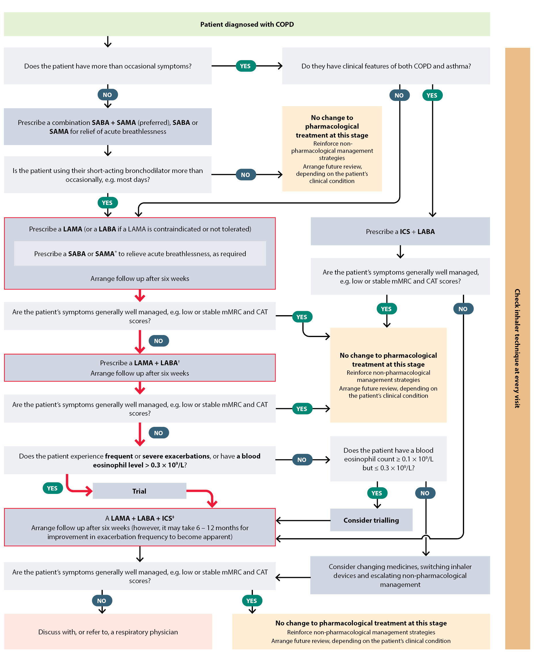
Figure 1. Suggested stepwise treatment for patients with COPD.4
Red pathway indicates prompt treatment escalation (recommended)
* SAMA and a LAMA should not be used concurrently
† A LABA + LAMA (single inhaler combination) is preferred, however, current Special Authority criteria for a LABA + LAMA single inhaler requires patients to be stabilised on LAMA treatment first
‡ A LABA + LAMA + ICS (single inhaler combination) is preferred — Special Authority criteria for a LABA + LAMA + ICS single inhaler requires patients to be on ICS + LABA or LABA + LAMA combination treatment, or multiple inhaler triple treatment (i.e. ICS + LAMA + LABA) first
CAT = COPD Assessment Test; COPD = chronic obstructive pulmonary disease, ICS = inhaled corticosteroid; LABA = long-acting beta2-agonist; LAMA = long-acting muscarinic antagonist; mMRC = Modified Medical Research Council Dyspnea Scale; SABA = short-acting beta2-agonist; SAMA = short-acting muscarinic antagonist.
All patients with symptomatic COPD require a bronchodilator
Short-acting bronchodilator treatment for acute dyspnoea
Almost all patients with symptomatic COPD will require a short-acting bronchodilator inhaler for acute symptom relief. However, prescribing a short-acting bronchodilator alone (i.e. without a long-acting medicine) for COPD is rarely indicated.4 This approach should only be considered in patients who report “very occasional” symptoms.4 If only a short-acting bronchodilator is appropriate, a combination short-acting beta2-agonist (SABA) + short-acting muscarinic antagonist (SAMA), e.g. salbutamol + ipratropium produces greater improvements in lung function and symptoms compared with either a SABA or SAMA alone.1 Escalation to a LAMA (with withdrawal of the SAMA; see below) is recommended if a patient reports regular use of a short-acting bronchodilator.4
Long-acting bronchodilator treatment for frequent symptoms
In most cases, a short-acting bronchodilator will not be sufficient to manage the patient’s symptoms. Patients with COPD who use their short-acting bronchodilator inhaler more than occasionally, i.e. on most days, should be prescribed a long-acting bronchodilator as well. New Zealand guidelines recommend prescribing a long-acting muscarinic antagonist (LAMA), e.g. glycopyrronium, tiotropium, or umeclidinium, first-line because of reduced exacerbation risk (compared to a long-acting beta2-agonist; LABA). A LABA should be selected only if a LAMA is contraindicated, not tolerated or the patient has features of both COPD and asthma (in combination with an ICS).4 Choosing a LAMA first-line is also practical, given prompt escalation is recommended in most patients and Special Authority criteria for a LAMA + LABA single inhaler requires patients to have trialled LAMA monotherapy.4
Be aware, a LAMA and a SAMA should not be used concurrently.4 For patients prescribed a LAMA, prescribe a SABA, e.g. salbutamol, terbutaline.4 For patients prescribed a LABA, prescribe either a SABA or a SAMA, e.g. ipratropium.4
Best Practice Tip: Frequent use of short-acting bronchodilators for acute symptom relief should prompt review of the patient’s treatment regimen, e.g. check inhaler technique and medicine adherence, consider escalation of treatment.
Prompt treatment escalation is recommended in most patients with COPD via two pathways
Persistent breathlessness add a second long-acting bronchodilator4
Escalate to dual combination treatment with a LAMA + LABA for patients with persistent symptoms, despite regular and correct use of a single bronchodilator medicine.4 Ideally, a single inhaler should be used to optimise adherence, however, patients initiated on a LABA will need to also trial a LAMA before meeting Special Authority criteria for a funded LAMA + LABA inhaler (Table 6).
Frequent or severe exacerbations add a second long-acting bronchodilator and consider ICS4
Consider adding an ICS to a LAMA + LABA dual combination inhaler treatment regimen in patients who have persistent symptoms and experience ongoing exacerbations or require hospitalisation.4 Patients with an eosinophilic pattern of COPD, i.e. blood eosinophil level > 0.3 × 10⁹/L, may be more likely to benefit from triple treatment, compared with dual LABA + LAMA treatment (see: “The role of eosinophils in COPD pathophysiology and management”).4 An ICS + LABA + LAMA single inhaler is preferred in patients who meet Special Authority criteria (Table 6), as this optimises treatment adherence.4
Triple treatment could also be considered for patients who use LAMA + LABA dual combination treatment but continue to report severe symptoms, e.g. persistent dyspnoea, exercise limitations, CAT score > 10 or have a blood eosinophil count ≥ 0.1 × 10⁹/L but ≤ 0.3 × 10⁹/L.1, 4
Options for patients in whom ICS is not recommended
The addition of an ICS is not recommended in patients with a blood eosinophil count < 0.1 × 10⁹/L who, despite the use of dual LAMA + LABA treatment, continue to experience dyspnoea (without exacerbations).1 In these patients, reassess inhaler technique and medicines adherence before changing medicines or switching inhaler devices and escalating non-pharmacological management, e.g. re-referral to pulmonary rehabilitation.1 Consider discussion with, or referral to, a respiratory physician.

Use of ICS treatment alone is not routinely recommended in patients with COPD as it has not conclusively been found to modify the long-term decline in the FEV1 or mortality risk (and it is an unapproved indication).1 Any potential reduction in exacerbation risk will likely be offset by the increased risk of pneumonia or other adverse effects.25
The role of eosinophils in COPD pathophysiology and management
COPD airway inflammation was conventionally thought to involve neutrophils and be associated with Th1/Th17 pathways.26 More recently, a pattern of disease involving eosinophils and type-2 inflammation, i.e. an eosinophilic phenotype, has been identified in a subset of people with COPD.1, 26 Eosinophils are granulocytic leukocytes that proliferate in response to type-2 inflammation mediators and are generally associated with asthma disease pathology.27, 28 Is it estimated that 20 – 36% of people with COPD have an eosinophilic pattern, depending on the definitions and thresholds used.1, 26 Older males with a higher BMI and a history of smoking are more likely to have this phenotype.26 People with an eosinophilic pattern of COPD have an increased risk of exacerbations and lung function decline, and are more likely to benefit from ICS treatment.26
Use blood eosinophil count and exacerbation history to determine ICS benefit
The presence of eosinophils in sputum is a marker of eosinophilic airway inflammation, however, measurement is not practical in primary care.28 Blood eosinophil counts correlate moderately with sputum eosinophil levels and are used as a surrogate to indicate the degree to which treatment with an ICS is likely to prevent future exacerbations.1, 28 The relationship between blood eosinophil counts and the protective (anti-inflammatory) effect of ICS treatment is continuous; ICS have no or very little benefit with a count < 0.1 × 10⁹/L, increasing to the greatest benefit with a count ≥ 0.3 × 10⁹/L.1
Blood eosinophil levels can also vary substantially over time due to circadian rhythm, seasonal changes, corticosteroid use, exercise, diet and infection.26 This variation is more common at elevated levels.1 Therefore, blood eosinophil counts guide treatment rather than being an absolute marker; thresholds are intended as an estimate. Exacerbation history is a stronger predictor of exacerbation risk; observational studies have shown that even one moderate exacerbation increases the likelihood of future exacerbations.1 An ICS would generally not be initiated without knowing the patient’s exacerbation history, i.e. number and severity.1, 25
Best Practice Tip: Avoid measuring blood eosinophils during an acute exacerbation if the patient has taken oral corticosteroids as they suppress eosinophil production and the result will not be informative.4 This is of particular importance if having a blood eosinophil level ≥ 0.3 × 10⁹/L in the past 12 months is the only way that a patient meets (or is able to meet) Special Authority criteria for funded treatment with a triple inhaler.
ICS + LABA treatment no longer routinely recommended
Reserve ICS + LABA inhalers for patients with features of both COPD and asthma.4 This shift is based on evidence that triple treatment, i.e. ICS + LABA + LAMA, is superior to ICS + LABA, if an inhaled corticosteroid is required.1 Patients with features of both COPD and asthma who continue to experience symptoms with correct use of their ICS + LABA should trial an ICS + LABA + LAMA single inhaler if they meet Special Authority criteria.4
Patients with COPD (but without clinical features of asthma or exacerbations) who are currently stabilised on ICS + LABA treatment can continue treatment.1 However, consider a switch to LABA + LAMA treatment if the patient’s symptoms worsen; further exacerbations indicate that escalation to triple treatment is required.1
Weigh potential benefits of long-term ICS treatment against the risk of adverse effects
ICS are potent anti-inflammatory medicines, and when used in combination with dual bronchodilator treatment, can reduce exacerbations by approximately 25% in some patients with COPD.25 However, their non-specific mechanism of action can also comprise the patient’s immune system, increasing the risk of respiratory infection and other adverse effects.25 The use of an ICS in patients with COPD is consistently associated with an increased risk of developing pneumonia; analysis of 19 randomised controlled trials found the risk of developing pneumonia increased by 41% with ICS use for one year or longer.25, 29 This is of particular concern in patients with other risk factors for pneumonia, e.g. current smoker, prior history of pneumonia, BMI < 25 kg/m2, severe airflow limitation.1
Discontinuation of ICS treatment in patients with COPD rapidly attenuates the elevated risk of pneumonia.5 A large, nested cohort study of more than 100,000 people with COPD treated with an ICS found a 37% decrease in the rate of serious pneumonia following ICS discontinuation.30
Other potential adverse effects associated with ICS include skin bruising, hoarseness and oral candidiasis; using a spacer with a metered dose inhaler and oral rinsing following actuation can help to reduce the risk of local fungal infection.1, 24 There is less robust evidence that ICS treatment is also associated with decreased bone density and increased fracture risk, increased risk of diabetes and reduced glycaemic control in people with diabetes, cataracts and mycobacterial infection, including tuberculosis.1 This increased risk of adverse effects is substantially lower than that associated with oral corticosteroids.1
Potential benefits of ICS likely outweigh the risks in patients with COPD and a blood eosinophil count ≥ 0.3 × 10⁹/L, however, the decision is less clear and needs to be individually weighed up in patients with lower blood eosinophil counts.25
When to consider withdrawal of ICS treatment
Consider withdrawal of ICS treatment if the patient:4
- Shows no evidence of benefit, i.e. no improvement in symptoms or fewer exacerbations
- Develops pneumonia or other ICS-related adverse effects
- Is clinically stable for 12 months and has had no exacerbations in that time
Check the patient’s blood eosinophil level prior to ICS withdrawal. A blood eosinophil count ≥ 0.3 × 10⁹/L increases the patient’s risk of exacerbations after stopping ICS and withdrawal may not be appropriate.4
Arrange follow-up four-to-six weeks after the patient stops ICS treatment to assess for worsening of symptoms or CAT score.4
Managing patients with clinical features of both COPD and asthma
Evidence suggests that people with features of both COPD and asthma (i.e. ACO) may experience worse outcomes than those with COPD alone, e.g. higher symptom burden, more frequent and severe respiratory exacerbations, poorer quality of life, however, these findings are not consistent across all studies.4, 31 People with mixed disease patterns are often excluded from clinical trials and treatment recommendations are therefore typically based on expert opinion.4 An action plan is guided by the patient’s dominant clinical features.4
ICS + LABA is recommended first-line for patients with features of both COPD and asthma; escalation to ICS + LABA + LAMA can be considered if patients continue to experience symptoms or exacerbations.4 Avoid LABA monotherapy in this patient group because it has previously been associated with a small but significantly increased risk of mortality in people with asthma.4, 32 There is currently insufficient evidence to support the use of ICS monotherapy or SMART/AIR therapy in patients with clinical features of COPD and asthma.4
Once stabilised, review patients with COPD regularly to identify disease progression, developing co-morbidities and any opportunities to improve management.1 Follow-up frequency is determined by the patient’s clinical condition; annual review is appropriate for patients with stable COPD whereas more regular review may be needed for those with more severe disease or co-morbidities.17
At every follow-up, discuss:4, 17
- Treatment adherence
- Symptom control (e.g. CAT score)
- Exacerbations – frequency and severity
- Smoking cessation support (if applicable)
- Vaccinations – check if due for influenza, pneumococcal or COVID-19
Depending on the clinical situation, additional investigations may also be required at some follow-up appointments:1, 4
- Measure oxygen saturation if appropriate, e.g. the patient has had a recent exacerbation or reports severe symptoms; this will support referral for long-term oxygen therapy in patients with advanced COPD
- Repeat spirometry if the patient has had a recent exacerbation or their clinical condition has deteriorated
- Consider arranging a chest X-ray and referral for non-acute respiratory assessment and chest CT if there has been a substantial deterioration in the patient’s clinical condition since their last review
The findings of this review may suggest escalation or reduction in the patient’s inhaled medicine regimen is required. However, check adherence to inhaler technique and non-pharmacological interventions before adjusting treatment.4 If the treatment regimen is modified, arrange a follow-up to evaluate the response after six weeks, using the CAT score and comparison to previous results to quantify benefit.4
Review any previously diagnosed co-morbid conditions.4 Regular follow-up also provides opportunities to assess the patient for developing conditions that adversely affect COPD, e.g. lung cancer, cardiovascular disease, anxiety, depression. When identified, a “treatable traits” approach is recommended.4 This strategy favours individualised disease management by addressing specific characteristics of respiratory disease and co-morbid conditions that contribute to the patient’s clinical status with the overall goal of improving their quality of life.33
A template for a four-step COPD consultation is available in Appendix 2 of the New Zealand COPD Guidelines: 2025 Update.
Keep cardiovascular disease, and particularly heart failure, in mind during follow-up
People with COPD have a higher risk of developing cardiovascular disease, including hypertension, ischaemic heart disease, atrial fibrillation and heart failure.1, 4 Heart failure in particular, requires extra consideration given its impact on COPD progression, as well as the overlap in symptoms and the potential for missed diagnosis (see: “The differential diagnosis of COPD”).20 COPD influences underlying cardiovascular dysfunction via systemic inflammation, pulmonary hypertension, impaired ventricular filling and ventricle wall remodelling.1, 20 These effects exacerbate heart failure symptoms and contribute to disease progression, i.e. pulmonary heart disease, including “cor pulmonale”.1, 20 At the same time, heart failure-associated volume overload and increases in left atrial pressure in different states, e.g. at rest, during exercise, lying supine, promote pulmonary congestion reducing lung compliance and worsening the discrepancy between ventilation and perfusion.20 Early diagnosis and optimising management of both COPD and heart failure is critical to slow disease progression and improve long-term outcomes in these patients.
For further information on the diagnosis and management of heart failure in primary care, see: https://bpac.org.nz/2025/heart-failure.aspx
When to discuss advance care planning for people with COPD
COPD is a progressive disease. Early discussions regarding changing treatment goals and advance care plans can be useful to open communication with patients and their family/whānau, with the goal of reducing decision burden and facilitating dignified end-of-life care. Clinical features that may prompt these discussions include patients with a FEV1 < 30% of predicted, those experiencing dyspnoea at rest, multiple exacerbations within 12 months, weight loss or cachexia or meeting criteria for oxygen therapy.4 A discussion about palliative care may also be appropriate in patients with severe symptoms and poor quality of life.4
Managing COPD exacerbations in primary care
Exacerbations are an acute worsening of COPD symptoms outside of expected day-to-day variation, e.g. increased dyspnoea, cough, sputum production or purulence (Table 7).1 Exacerbations are associated with increased airflow obstruction, disease progression, hospitalisation and mortality.1 Triggers include viral or bacterial infection, changes in ambient temperature and environmental pollutants.34
Exacerbations can often be managed in the community, however, prompt identification and treatment is critical as even patients with mild COPD can experience deterioration in lung function following these events.4 Consider performing an ECG and arranging appropriate investigations (e.g. BNP) and imaging (e.g. chest X-ray) to rule out other causes of symptoms and identify potential complications, e.g. heart failure, arrhythmias, pulmonary embolism.4 Provide education to both the patient and their family/whānau to ensure they can take the appropriate steps and seek medical attention early. Develop an exacerbation plan with the patient and regularly review this as part of ongoing follow-up.

Acute referral to hospital should be considered in the following situations:4
- Insufficient or no response to medical management
- A sudden worsening of symptoms
- Confusion or drowsiness
- Cyanosis and peripheral oedema
- Low oxygen saturation (SpO2 < 85 – 90%)
- Co-morbidities, e.g. heart failure, newly occurring arrhythmias
- Living circumstances are not appropriate, e.g. limited home support, lack of telephone, transport or distance to hospital
Table 7. Classification of COPD exacerbation severity.4 Adapted from New Zealand COPD Guidelines: 2025 Update.
| Classification |
Features |
| Mild-to-moderate |
More short of breath than usual
Unable to speak in sentences
Wheeze often present
Some indrawing of the chest/neck, e.g. tracheal tug
SpO2 near usual level
Normal level of consciousness |
| Severe |
Very short of breath
Only a few words per breath
Severe indrawing of the chest/neck, e.g. tracheal tug
Tripod positioning, i.e. leaning forward, resting elbows or hands on knees/surface to relieve dyspnoea
SpO2 well below usual level
Agitation |
| Life-threatening/imminent respiratory arrest |
Extremely short of breath
Unable to speak
Wheeze may not be present
Indrawing of the chest/neck can be absent
SpO2 rapidly decreasing
Severe agitation or reduced consciousness |
Pharmacological management of COPD exacerbations

A SABA inhaler, e.g. salbutamol, is recommended first-line in patients experiencing an exacerbation.4 A SABA + SAMA combination inhaler is also appropriate, e.g. salbutamol + ipratropium (unless the patient is prescribed a LAMA).4 Ideally, patients prescribed pressurised metered dose inhalers would use a spacer. Advise patients to continue maintenance bronchodilator (and ICS) treatment during an exacerbation.

Oral antibiotics can be considered for patients with dyspnoea and other clinical features suggestive of bacterial infection, including fever, purulent sputum, increased sputum volume or elevated inflammatory markers, e.g. C-reactive protein > 50 mg/L (if results are available).4 See Table 8 for recommended antibiotic options. Sputum culture is not routinely requested in primary care, however, may be appropriate in patients experiencing recurrent exacerbations or an insufficient response to empiric antibiotics, or under the direction of a respiratory physician.4, 35
Table 8. Antibiotic options for COPD exacerbations in primary care.4, 36
| Antibiotic |
Dose |
| Amoxicillin |
1,000 mg, three times daily, for five days |
| If penicillin allergy: |
| Doxycycline |
200 mg, on day one (loading dose), followed by 100 mg, twice daily, on days two to five |
| If past antibiotic treatment has been unsuccessful or resistant organisms have been previously isolated: |
| Amoxicillin + clavulanic acid |
625 mg, three times daily, for five days |

Short-term oral corticosteroids should be considered in moderate to severe exacerbations.4 Prescribe 40 mg prednisone, once daily, for five days.4 Tapering is not usually required, but should be considered if treatment lasts longer than 14 days,5 or other factors are present, e.g. history of adrenal insufficiency. Do not prescribe long-term oral corticosteroids for COPD exacerbations as they increase the risk of adverse effects, e.g. pneumonia, osteoporosis, and mortality.1, 5
Arrange post-exacerbation follow-up
Patients may take up to six weeks to fully recover from an exacerbation, and in some cases, their pulmonary function may not return to their previous level.1 A review of the patient’s COPD management, including current medicines, inhaler technique, adherence and non-pharmacological interventions, is recommended following every exacerbation.4, 34 Refer the patient for pulmonary rehabilitation, unless they have completed the course within the past 12 months or it is contraindicated (e.g. recent cardiac event, unstable angina or medical condition that restricts movement such as severe arthritis).4 Spirometry could also be considered to reassess lung function.