Classification and aetiology of urticaria
Urticaria is the term used to describe a group of skin conditions, characterised by the presence of wheals. Approximately
one in five people experience urticaria (commonly referred to as hives) at some stage in their life.1,2 In
many cases, a specific trigger for the urticaria is not found. In rare cases, urticaria may be a sign of systemic disease,
such as an autoimmune condition.
The two main classifications of urticaria are:
- Ordinary (spontaneous) urticaria – which can be acute or chronic
- Physical urticaria
Acute urticaria describes "one-off" outbreaks and recurrent episodes occurring over a period of less than six weeks.
It is the most common type of urticaria, and is more frequently seen in children and young adults.1,3 It
is estimated that 20 – 30% of cases of acute urticaria in infants and young children develop into chronic urticaria.4 Approximately
50% of cases of acute urticaria are idiopathic, i.e. a specific trigger is not identified.3
Chronic urticaria describes episodes of urticaria which occur over a period longer than six weeks. In rare cases urticaria
may persist for a lifetime, but this is more common in cases of physical urticaria .5 Approximately 30% of
patients presenting in primary care with urticaria will have chronic urticaria.6 Chronic urticaria occurs
more frequently in adults, and in women (approximately 60% of cases).1 It is estimated that in 40% of people
with chronic urticaria, there is evidence of an autoimmune process, and in 20% there is evidence of a physical stimulus,1 although
a specific cause is often not found.
Physical urticaria occurs in a localised area after contact with a physical stimulus. Individual episodes usually
resolve within a two hour period, but physical urticaria often persists as a chronic, recurring condition.3 Dermatographism
(skin writing) is the most common form of physical urticaria, triggered by firm stroking or scratching of the skin,
or contact with clothes or other objects (Figure 1).3
Other types of physical urticaria include;
- Contact urticaria – absorption of substances through the skin or mucous membranes
- Cholinergic urticaria – sweating, e.g. after exercise or exposure to heat
- Delayed pressure urticaria – sustained pressure to a site on the body, e.g. on the buttocks after sitting
- Cold urticaria – most frequently caused by swimming in cold water or exposure to cold wind (Figure 2)
- Solar urticaria
- Vibratory urticaria
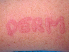
Figure 1: Dermatographism
Images provided by DermnetNZ |
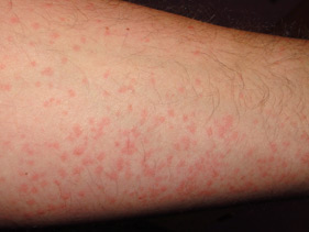
Figure 2: Cold urticaria |
Most cases of urticaria are non-allergenic
Most cases of urticaria are not caused by allergy but are the result of histamine being released by direct mast cell
degranulation (i.e non-IgE mediated).
Examples of causes of non-allergenic urticaria include:2
- Infection – bacterial (e.g. Helicobacter pylori, Mycoplasma pneumoniae), viral (e.g. infectious mononucleosis, viral
hepatitis), parasitic (e.g. Giardia) or fungal (e.g. Candida)
- Medicines – especially opiates, aspirin and non-steroidal anti-inflammatory drugs (NSAIDs)
- Non-allergenic contact with topical compounds, food preservatives, raw meat or vegetables
- Non-allergenic food reactions to compounds such as alcohol, salicylates in fruit or from bacterial decomposition
(food poisoning)
- Hypersensitivity to physical stimuli such as scratching, friction from clothing or other objects, light, heat, cold,
water or vibration
- Autoimmune conditions such as systemic lupus erythematosus and autoimmune thyroid disease
Allergy-induced urticaria
Allergy induced urticaria is most common in people with a history of atopy.
Examples of causes of allergenic urticaria include:2
- Medicines, e.g. antibiotics
- Food allergy, e.g. fish, eggs or nuts
- Insect stings, e.g. wasp, bee
- Contact allergens, e.g. latex or cosmetics
Clinical history and examination
Clinical history and physical examination are usually sufficient to diagnose urticaria. A specific cause is identified
in approximately one-half of patients with acute urticaria and one-quarter of patients with chronic urticaria.3,7
Clinical history
The clinical history should cover:
- Frequency, size, distribution and duration of the lesions – to determine type of urticaria
- Recent consumption of new or unusual food or medicines, recent infections, or participation in or exposure to new
activities, locations or products or chemicals – to determine potential triggers
- Occupational exposure to chemicals or inhalants – to determine potential long-term triggers
- History of similar episodes and response to treatment
- Personal and family history of atopy – more likely to be allergy-induced urticaria
Physical examination: clinical features of urticaria
An episode of urticaria is identified by highly pruritic, well-defined, pink-to-red wheals, often with a pale centre
(Figure 3), which usually last no more than 48 hours and leave no remaining marks. The lesions may occur anywhere on
the skin and can range in size, from a few millimetres to centimetres, and vary in shape, forming round, oval, annular
(ring) (Figure 4), serpiginous (wavy), gyrate (circular, coiled) or targetoid (target pattern) plaques. The lesions
may also merge to form large geographic or giant patches (Figure 5). The surface skin remains smooth. The presentation
of urticaria is similar in both children and adults.
Approximately 40% of people with urticaria also have signs of angioedema.1 Angioedema involves the deeper
epidermis and subcutaneous tissues and most frequently affects the eyes, mouth, throat, tongue, hands and feet. Angioedema
without urticaria is rare and can be life-threatening if the larynx is involved. Further discussion of this condition
is outside the scope of this article.
Further examination should be guided by the clinical history. Dermatographism can be tested for by stroking the skin
firmly and looking for linear wheals occurring within a five minute period. The application for several minutes of an
ice cube, heat, pressure or water may rule out other forms of physical urticaria.
In some cases, examination may be necessary for underlying conditions that may precipitate urticaria, such as:
- Bacterial or fungal infections of the skin
- Autoimmune thyroid disease – may be indicated by an enlarged thyroid
- Connective tissue diseases – may be indicated by joint swelling or tenderness or oral ulceration, e.g. rheumatoid
arthritis, systemic lupus erythematosus
- Liver disease/dysfunction – may be indicated by tenderness on palpation of the liver or jaundice, e.g. cholestasis
can cause pruritus and acute urticaria can be an early sign of hepatitis A, B and rarely C8
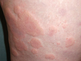
Figure 3: Classical whealing |
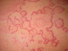
Figure 4: Annular pattern |
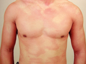
Figure 5: Giant urticaria |
| Images provided by DermnetNZ |
Differential diagnosis
There are a large number of conditions (some of them rare) which may cause symptoms similar to urticaria. The transient
and pruritic nature of lesions is one of the most distinctive aspects of urticaria, but pruritus is sometimes absent.
Angioedema is also more likely to be associated with urticaria than other skin conditions.
If the signs and symptoms are not typical of urticaria, other diagnoses that may be considered include:
- Atopic dermatitis – usually highly pruritic, but can be distinguished from urticaria by the lack of transitory wheals,
excessively dry skin and other skin surface abnormalities, strongly associated with personal or family history of atopy
- Contact dermatitis – can be distinguished from urticaria by a lack of transitory wheals and the presence of skin
surface changes such as blisters, dryness and peeling
- Fixed drug eruptions – tender, well defined, round or oval patches, often with central blistering that generally
occur in the same place on the body each time a specific medicine is taken
- Erythema multiforme – an acute, and at times recurring, hypersensitivity to a variety of causes including infections
and medicines. Lesions are usually present on the face and distal limbs and can last for up to seven days.
- Bullous pemphigoid – a chronic, autoimmune condition, which usually affects elderly people. Characterised by erosions
and tense bullae filled with clear, cloudy or blood-stained fluid, most frequently occurring in body folds.
- Urticarial vasculitis – characterised by wheals that resemble urticaria, but last longer than 48 hours and often
leave bruising and areas of increased pigmentation as they resolve
- Papular urticaria – urticated pruritic papules at the site of insect bites, common in young children and in people
who have travelled.
Laboratory investigation of urticaria
Laboratory testing is not indicated for patients with acute urticaria as the diagnosis is usually clinical.
In patients with chronic urticaria, testing does not usually help to establish a cause, direct management or improve
patient outcomes.9 In a study of 356 patients with urticaria referred for allergy and immunology evaluation,
only one patient benefited from a change in management due to testing and only 319 (17%) of the 1872 tests ordered had
abnormal findings.10
Laboratory testing may be useful in selected patients with chronic urticaria, e.g. if an underlying condition is suspected,
they have failed to respond to treatment, or if the condition is severe.9 The choice of investigations should
be guided by positive findings from the clinical history and physical examination. Discussion with a dermatologist may
also be helpful.
The following investigations may be appropriate for specific clinical circumstances:
Skin prick testing may be considered when an allergic cause for the urticaria is suspected and confirmation would be
useful for management, e.g. if avoidance measures are being considered. Skin prick testing should not be performed routinely.
Skin prick testing may not be reliable in older adults and children aged under two years should be referred to an allergy
clinic for testing as the results may be difficult to interpret. Skin prick testing in pregnant women should only be
requested if the benefits outweigh the risks, as in rare cases it can cause uterine contractions.11
Serum allergen-specific IgE testing is second-line to skin prick testing when skin prick testing is unsuitable or unavailable.
 For further information see: "Appropriate use of allergy testing in
primary care", Best Tests (Dec, 2011)
For further information see: "Appropriate use of allergy testing in
primary care", Best Tests (Dec, 2011)
Full blood count may indicate an allergy or an intestinal infection if the eosinophil count is elevated. Neutropenia
may suggest an autoimmune or viral cause, while neutrophilia may be caused by a bacterial infection. Acute viral infections,
e.g. Epstein-Barr virus, or autoimmune thyroiditis may cause a high lymphocyte count.
Thyroid antibody testing may be useful following discussion with an appropriate specialist, if a thyroid autoimmune
disorder is suspected. Chronic autoimmune urticaria is associated with antithyroid antibodies in approximately one-quarter
of cases.3
Skin biopsy (3 mm punch biopsy) is only rarely required, if urticarial vasculitus is suspected or when the diagnosis
is uncertain. Atypical features of urticaria include pain or burning rather than pruritis, complete non-response to anti-histamines,
wheals persisting for longer than 48 hours, or not fully resolving, with remaining hyperpigmentation.
Best Practice tip: Before contacting a dermatologist, take anatomic
views and close-up digital images of the patient's skin lesions. Emailing good quality clinical images may assist the
discussion, particularly if the patient's clinical signs are intermittent.
Treatment for urticaria
Acute urticaria generally resolves over a short period of time, however, chronic urticaria can persist for months
or even years (particularly physical urticaria). This can be frustrating for both patient and doctor, especially when
there is no known cause.
In a study of 220 patients with chronic idiopathic urticaria, it was found that after one year:6
- 47% were symptom-free
- 60% with ordinary urticaria and angioedema were symptom free
- 39% with ordinary urticaria only were symptom free
- 16% with physical urticaria were symptom free
Management is focused on avoiding triggers where known, and using medicines for symptom relief.
Avoidance strategies
When the clinical history does not reveal an obvious cause for the urticaria, an avoidance strategy for potential
triggers may be considered.
Patients can be advised to stop any non-essential medicines, herbal supplements or topical preparations. In particular,
aspirin, codeine and non-steroidal anti-inflammatory drugs (NSAIDs) may contribute to wheal formation, even when they
are not the primary cause of the eruption. If symptoms resolve (or do not recur), medicines/products can be reintroduced
sequentially, if necessary, and the patient should report any return of symptoms.
Dietary investigations rarely identify a specific trigger for chronic urticaria, and are not necessary in cases where
symptoms can be easily controlled with oral antihistamines. However, if the patient wishes to, a food diary may be used
to record and eliminate suspected triggers. Particularly motivated people may try a narrow diet of rice and a single
source of protein for two weeks, while discontinuing all antihistamines. Foods can then be slowly reintroduced and reactions
noted in the food diary.1
Pharmacological treatment
Introduction of medicines for the treatment of urticaria should be considered in the following order:
- Commence non-sedating oral antihistamines
- Add conventional sedating oral antihistamines and/or H2 receptor antagonists
- Add tricyclic antidepressants
- Add oral corticosteroids - only for patients with severe acute urticaria
Non-sedating oral antihistamines are the first-line pharmacological treatment for both acute and chronic urticaria
due to their effectiveness and relative lack of anticholinergic and central nervous system effects. Although referred
to as "non-sedating", these medicines may still cause sedation at usual doses in some patients. In New Zealand cetirizine
and loratadine are fully-funded (see Table 1 for recommended doses). Individual response to antihistamines may be variable,
however, cetirizine is thought to be the quickest acting, therefore may be trialled first.2
Table 1: Recommended doses for fully-funded, non-sedating antihistamines available in New Zealand13,14,15
| Antihistamine |
Adult dose |
Child dose (6 –12 years) |
Child dose (2 – 6 years) |
| Cetirizine |
10 mg, once or twice daily* |
10 mg, once daily or in divided doses |
5 mg, once daily or in divided doses |
| Loratadine |
10 mg, once or twice daily* |
> 30 kg: 10 mg, once daily
< 30 kg: 5 mg, once daily |
5 mg, once daily |
* Although the maximum dose in the New Zealand medicine datasheet is 10 mg, this medicine is often used (and
required) in higher doses, without any reports of adverse effects, in order to successfully manage urticaria
12,16
Oral antihistamines may be taken on an "as-required" basis, due to their rapid onset of action, but may be more effective
when taken daily. The recommended maximum adult dose of cetirizine and loratadine is 10 mg per day, however, European
guidelines recommend non-sedating antihistamines be prescribed at up to four times the standard dose (i.e. cetirizine
or loratadine 40 mg daily) before second-line medicines are considered as adjunctive treatment.12
Sedating oral antihistamines are rarely used as a monotherapy for urticaria, but can be used in combination with non-sedating
antihistamines. These medicines may be useful for patients with nocturnal symptoms that prevent sleep. Promethazine
(fully funded) is a suitable choice and can be prescribed at the following doses:13,14
- Adults; 25 – 75 mg, at night
- Children aged five to ten years; 10 – 25 mg, at night
- Children aged two to five years; 5 – 15 mg, at night
H2 receptor antagonists such as ranitidine or famotidine, when used in combination with antihistamines,
may be of benefit to some people with chronic urticaria as 15% of histamine receptors in the skin are H2-type.3 These
medicines are not recommended as monotherapy because their ability to reduce pruritus is limited and there is little
clinical evidence of their effectiveness.
Tricyclic antidepressants have histamine receptor antagonist activity and may be especially useful
in treating chronic urticaria, in combination with non-sedating antihistamines. Due to its sedating properties doxepin
(30 – 50 mg) is an appropriate treatment for nocturnal symptoms. Amitriptyline (10 – 50 mg) may also be effective.
Oral corticosteroids may be added for people with severe acute urticaria. The recommended dose for
adults is 20 – 40 mg daily, or for children 1 mg/kg daily, maximum 40 mg, tapering to the lowest effective dose over
the course of two to five days.13 Corticosteroids are nearly always inappropriate in people with chronic
urticaria as long-term use should be avoided.
N.B. Topical corticosteroids are not useful in the treatment of urticaria and may cause adverse effects with longer-term
or higher-potency use, e.g. skin atrophy. Topical antihistamines are also not effective for treating urticaria and are
not recommended due to the risk of sensitisation and resulting contact dermatitis.17
Cooling preparations containing 0.5 – 1% menthol in a cream or lotion base, e.g. cetomacrogol cream, may provide symptom
relief. The use of cool damp cloths, reduction of night-time heating and tepid showers may also be useful.
Referral for specialist treatment may be considered if the diagnosis is uncertain or where symptoms are severe and
poorly controlled. A number of further treatment options are available including immunosuppressants, e.g. cyclosporin,
and leukotriene receptor agonists, e.g. montelukast. If a complex drug or food trigger is suspected then consider referral
to an immunologist. Phototherapy using ultraviolet B radiation reduces the number of mast cells in the upper dermis,12 and
may be effective in reducing symptoms in cases of physical urticaria that are resistant to antihistamines.18 Patients
can be referred to a dermatologist for this treatment.
Best Practice tip: A standard treatment regimen for urticaria – begin with
cetirizine, if symptoms are not controlled, add promethazine 25 mg at night and raniditine 300 mg during the day. This
will settle symptoms for most people. If symptoms still persist, add in a tricyclic antidepressant.