Fungal infection accounts for approximately half of all nail disease
Fungal infection of the nails or onychomycosis accounts for about 50% of all nail disease. It becomes more common with
increasing age and mostly affects toenails (80% of cases). Other risk factors for onychomycosis include: nail trauma,
frequent immersion in water, occlusive footwear, athletes foot, diabetes mellitus, immunosuppression and smoking.1,2,3
Dermatophytes are the most common cause of fungal nail infection
In 90% of cases onychomycosis is caused by a dermatophyte, mostly Trichophyton rubrum. Other causes of infection
include yeasts; mainly Candida infection of the fingernails, which is often accompanied by paronchynia (inflammation
of the proximal nail fold).1 Non-dermatophyte moulds (Fusarium, Scopulariopsis and Scytalidium)
account for about 2–3% of fungal nail infections.2,4
Distal and lateral subungual onychomycosis is the most common morphology of fungal nail infection
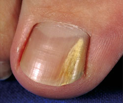 |
| Fig 1: Typical lateral onychomycosis (T. rubrum) |
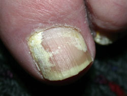 |
| Fig 2: Extensive superficial white onychomycosis (T. interdigitale) |
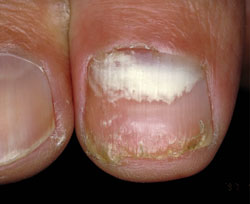 |
| Fig 3: Proximal onychomycosis (T. interdigitale) |
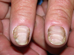 |
| Fig 4: Typical onychomycosis and paronychia due to C. albicans |
Most cases of onychomycosis are characterised by thickening of the nail, discolouration (ranging from white to black)
and onycholysis (separation of the nail from the nail bed).1,5
There are different morphological types of onychomycosis, the most common being distal and lateral subungual onychomycosis
(Figure 1), usually caused by dermatophyte infection.1,5 This begins in the distal or lateral part of the nail
plate and spreads proximally under the nail.
Superficial white onychomycosis (Figure 2) is also mostly caused by dermatophyte infection (usually Trichophyton
interdigitale) and accounts for about 10% of onychomycosis. It presents as small, white, powdery patches on the
surface of the nail.2,6
Proximal onychomycosis (Figure 3) is the least common type of onychomycosis and usually presents in patients who are
immunosuppressed (e.g. HIV), have diabetes, or in those with peripheral vascular disease.1,2 This begins as
discolouration of the proximal end of the nail.
Candidal onychomycosis (Figure 4) is often associated with paronchynia and most often occurs in the fingernails of people
who frequently immerse their hands in water.1,2
Laboratory diagnosis is recommended before any treatment is started
Laboratory diagnosis is recommended before starting treatment because other conditions (see differential diagnoses below)
can present similarly to onychomycosis, particularly psoriasis. Also, treatment of onychomycosis is lengthy, may have
adverse effects and can be expensive.1,2,3
Diagnose onychomycosis with microscopy and culture of nail clippings7
Microscopy of a sample of affected nail plate can identify fungal elements and culture determines the causative organism.8
In most cases patients will be sent to the laboratory for collection of samples, however in some cases (e.g. rural practice)
the doctor may collect the sample. Nail clippings (chiropody clippers may be the best tool to use) from the diseased part
of the nail and curettings of subungual debris should be taken. If superficial white onychomycosis is suspected, a scalpel
can be used to scrape the surface of the nail to obtain a sample.1,2
It is best to provide the laboratory with a generous amount as there are usually few fungi in a typical specimen. If
necessary, delay the investigation to allow the nail to grow longer.2,8
Problems with laboratory tests – false negatives and delayed results
Accurate microscopy of specimens depends on the skill of the laboratory personnel. The false negative rate can be 30–40%.5 Mycological
culture increases the sensitivity but results may take several weeks because dermatophytes are slow growing. A culture
plate is incubated for four weeks before it is declared negative.9
Differential diagnoses1,2
Psoriasis affecting the nail – pitting, onycholysis, discolouration, thickening and irregular ridging. Look for psoriatic
plaques on typical sites (scalp, ears, elbows, knees and flexures).
Onychogryphosis – thickening and distortion of the nails, most often the big toe. This is more common in elderly people.
Other differential diagnoses include – lichen planus, nail trauma, squamous cell carcinoma and malignant melanoma and
nail dystrophy caused by systemic disease.
Treatment options
Treatment may not be necessary for everybody and may be inappropriate for elderly people
For patients who do not have troublesome symptoms, the decision to treat can be based on the patient’s choice, after
they have been informed of risks and benefits of treatment.
Patients should be informed about treatment including:1
- Nail may not look completely normal, even after treatment
- Treatment with oral antifungals is only successful in about 70-80% of cases (and clinical trials usually exclude people
aged over 60 years) and can relapse.8 Topical treatment is less effective.10
- Length of treatments: three months for oral medication and up to two years for topical medication
- Potential adverse effects and interactions with oral treatment
Treatment with oral antifungals may not be appropriate for elderly people, or people taking multiple medicines, as there
is an increased risk of adverse effects and interactions.2
Treatment should be considered for those at risk of complications
People with diabetes, peripheral vascular disease or connective tissue disorders are at higher risk of complications
such as secondary bacterial infections (e.g. cellulitis). In these people treatment should be considered. However, treatment
is less likely to be successful in these patients, who also have higher rates of drug-induced complications.1,11
Pharmacological treatments
Oral antifungals (terbinafine, itraconazole) and topical antifungals (ciclopirox, amorolfine) are available for the
treatment of onychomycosis.
Oral treatment is more effective than topical therapy and is recommended for most people in whom treatment is appropriate
Oral antifungal treatment is more effective than topical treatment except for cases of superficial white onychomycosis
or in a few cases of minor distal onychomycosis. Oral therapy is considered first line for most patients who opt for treatment.8
Terbinafine is first line for dermatophyte infection
Terbinafine and itraconazole are both effective for treating dermatophyte infection however terbinafine is more
effective and is considered first line.2,8,11
In adults, 250 mg terbinafine is given once daily for an initial course of six weeks for typical fingernail infection,
or 12 weeks for toenail infection.
Gastrointestinal effects such as dyspepsia, nausea and diarrhoea, skin reactions (morbilliform rash, urticaria) and
taste disturbance are the most common adverse effects associated with terbinafine.1,2
Rarely, serious skin reactions (e.g. Stevens-Johnson syndrome, toxic epidermal necrolysis, drug hypersensitivity syndrome
and angioneurotic oedema) and haematological disorders have occurred with terbinafine therapy.2 Psoriasis may
be aggravated by terbinafine treatment.2
Oral terbinafine is not recommended for people with liver disease. Patients taking terbinafine should be advised to
promptly report any symptoms that may suggest liver toxicity such as anorexia, nausea, vomiting or fatigue.12 In
elderly or unwell people, liver function and blood count should be monitored at baseline and then after four to six weeks
of treatment.
Terbinafine interacts with a small number of drugs (Table 1).
Itraconazole is the treatment of choice for onycho-mycosis due to candida infection
Itraconazole is the most effective treatment for onychomycosis due to candida infection.8,11 Terbinafine can
be used but is less effective.1
For the treatment of onychomycosis in adults, 200 mg itraconazole is taken twice daily for seven consecutive days of
the month, for two months for fingernails and three months for toenails (although candidal onychomycosis is much less
common in toenails).8 Alternatively, 200 mg itraconazole can be taken once daily for three months. There is
no evidence that continuous or intermittent regimens produce significantly different cure rates or adverse events.13
Adverse effects associated with itraconazole also include gastrointestinal effects and skin reactions as well as reversible
increases in hepatic enzymes. Uncommon adverse effects include hepatotoxicity, severe skin conditions, nervous system
disorders (peripheral neuropathy, headache, dizziness) and congestive heart failure.2
Itraconazole should not be used in patients with congestive heart failure or in those with liver disease or raised liver
enzymes.
LFTs should be monitored at baseline and after four to six weeks of treatment for courses lasting more than one month.1
Itraconazole is an inhibitor of CYP3A4 and has a number of significant drug interactions (Table 1).
Table 1: Interactions with antifungals 2,3,14
| Drug or class |
Terbinafine |
Itraconazole |
| Benzodiazepines |
|
Concurrent use of midazolam or triazolam is contraindicated, risk of excessive or prolonged sedation |
| Statins |
|
Avoid concomitant use as rhabdomyolysis has been reported |
| Rifampicin |
Decreased plasma levels of terbinafine possible |
|
| Warfarin |
Bleeding events reported rarely, however avoiding concurrent use is not necessary |
Increased risk of bleeding, increased INR monitoring may be required |
| Calcium channel blockers |
|
Increases the plasma level of felodipine which may increase its adverse effects, particularly ankle
and leg oedema |
| N.B. This is not a comprehensive list of interactions |
Topical treatment may be suitable for superficial infection and for those unable to take oral antifungal therapy
Topical therapies are less effective than oral therapies but may be useful for superficial white onychomycosis or in
early distal and lateral subungual onychomycosis.11,15 Topical therapy can also be used where a patient is
unable or unwilling to take oral antifungals.11 Compliance with topical treatments can be an issue as they
require application for extended periods, e.g. 6 to 12 months.
Amorolfine and ciclopirox nail lacquers are available topical antifungals
Amorolfine 5% (Loceryl) and ciclopirox 8% (Batrafen) are two topical antifungals available in New Zealand. While
there is limited evidence that ciclopirox modestly improves symptoms of onychomycosis compared with placebo, there is
a lack of evidence examining effectiveness of amorolfine.16,17
Amorolfine is applied to the affected nail twice weekly until infection is resolved, usually six months
for fingernails and 9 to 12 months or longer, for toenails. The nail needs to be filed, cleansed and de-greased before
application.1,18 A transient burning sensation may occur after application of nail lacquer.1
Ciclopirox is applied to the affected nails every second day for the first month, then application
is reduced to twice weekly for the following month, and then reduced to once weekly for up to six months or longer. The
nail lacquer should be removed with nail vanish once weekly and the nail filed.18 Irritation and pruritus may
occur after application.
Both products are available over-the-counter as pharmacist-only-medicines or on prescription. There are part charges
for both and these products may be too expensive for some patients.
Measure treatment response as nail grows
To determine if treatment is effective, photograph the nail, or make a groove with a nail file at the proximal end of
the infected area. The infection should not progress proximal to this groove if treatment is effective. The groove may
need to be redefined over time because it can take 12 months or longer for a big toenail to grow out.2
All people, whether they are being treated with medicines or not, should be provided with lifestyle advice
Foot care advice is integral to the treatment of onychomycosis and may lessen the discomfort of the infected nail(s).2,3,15
Advise patients to:2,3,15
- Keep feet cool and dry by wearing cotton socks and breathable footwear
- Trim nails and file down hypertrophic nails
- Avoid high heels and narrow toed shoes to prevent nail trauma
- Recognise and treat athletes foot if present
- Wear footwear in communal showers
Treatment failure or relapse
If initial treatment fails or infection recurs, the first step is to confirm mycology.3 Earlier positive
culture may have been secondary infection of nail dystrophy due to another cause. In addition, check adherence and if
treatment is required, an alternative drug, a combination of oral and topical treatment or nail avulsion may be considered.
However, there is little evidence that nail avulsion increases cure rates.2,11
Images contributed by NZ DermNet, the website of the
New Zealand Dermatological Society.