This item is 7 years and 0 month old; some content may no longer be current.

How to use fluorouracil and imiquimod for non-melanoma skin cancer in a general practice setting

Fluorouracil and imiquimod creams are fully subsidised topical treatments, suitable for some patients with non-melanoma skin cancers; Special Authority approval is no longer required for subsidy. Their place in treatment depends on the type, severity and location of the lesion(s), as well as the expertise and experience of the prescribing clinician (in terms of other treatments which can be offered) and the patient’s preference.

0 comments
save
share
feedback
Log in

Key concepts

  • Fluorouracil and imiquimod creams are topical treatments that may be used to treat some non-melanoma skin cancers, usually second-line to surgical excision and/or cryotherapy
  • These medicines work by destroying cancerous cells in the skin, resulting in a local reaction including erythema and erosion, followed by re-epithelisation of the skin
  • Fluorouracil and imiquimod may be appropriate for the treatment of actinic keratoses, superficial basal cell carcinoma and squamous cell carcinoma in situ
  • Treatment regimens vary depending on the type of lesion, but fluorouracil and imiquimod creams are typically applied daily or several times a week, for four to 12 weeks or longer

Fluorouracil and imiquimod creams are fully subsidised topical treatments, suitable for some patients with non-melanoma skin cancers; Special Authority approval is no longer required for subsidy. Their place in treatment depends on the type, severity and location of the lesion(s), as well as the expertise and experience of the prescribing clinician (in terms of other treatments which can be offered) and the patient’s preference.

Surgical excision is the recommended first-line treatment for non-melanoma skin cancers in the majority of cases.1 Fluorouracil 5% and imiquimod 5% creams may be a treatment option for superficial lesions, where surgical excision or other treatments such as cryotherapy are not practical or desirable by the patient. Additional treatment options for non-melanoma skin cancers, e.g. ingenol gel (not subsidised – refer to the New Zealand Formulary ) and photodynamic therapy (using photosensitising drugs activated by specific kinds of light to target cancerous cells), may also be offered to patients.

Fluorouracil and imiquimod can be effective treatments for non-melanoma skin cancers, when used appropriately. When prescribing these medicines, ensure that patients understand how, where and when they should be applied, and what precautions to take when handling, storing and disposing of the medicine. It is important that patients avoid excessive sun exposure, especially with fluorouracil treatment.

N.B. This article does not cover the detection and diagnosis of non-melanoma skin cancers, the use of other treatments, such as cryotherapy, or the management of melanoma skin cancer. For further information on skin cancers, see: www.bpac.org.nz/BPJ/2013/December/skincancer.aspx

Actinic (solar) keratoses

These lesions, which are usually flat, scaly and non-pigmented, develop on skin damaged from ultraviolet (UV) light exposure. Actinic keratoses are benign but can progress to invasive malignant disease if left untreated.2 Surgical excision is not routinely performed, due to the nature of the lesions. However, some actinic keratoses may be hypertrophic, in which case shave excision can be performed.2 Biopsy is not usually necessary for an isolated lesion of typical appearance, but should be considered for patients with recurrent lesions or if the diagnosis is unclear.2 If the extent of actinic keratoses makes treatment impractical, long-term surveillance is essential for early diagnosis of malignancy.

Actinic keratoses usually respond well to cryotherapy, fluorouracil and imiquimod creams, ingenol gel and photodynamic therapy. A combination of methods may also be used.2

Cryotherapy is the usual treatment of choice for isolated and hyperkeratotic actinic keratoses. The outcome depends on the experience of the clinician performing this procedure, freeze time and the number of applications.2 The main disadvantage of cryotherapy is that only visible lesions are targeted. It is not uncommon for further lesions to emerge in the same area over time.2

Fluorouracil or imiquimod creams are effective treatments for flat actinic keratoses (Figure 1) , and are associated with a higher rate of long-term clearance of lesions at the treatment site than cryotherapy.2 A study found that one year after treatment, 73% of patients that used imiquimod and 33% of those who used fluorouracil had maintained clearance of actinic keratoses in the treated area compared to 4% of those who underwent two cycles of cryotherapy.3 Inflammation, erosion and pain associated with both fluorouracil and imiquimod may, however, mean that topical treatment is poorly tolerated and not able to be used for the necessary duration to achieve optimal results.2

Both fluorouracil and imiquimod can be applied to discrete lesions or applied to a wider affected area (field treatment). Field treatment can result in the emergence of lesions which were previously sub-clinical, but this is considered to be evidence of treatment efficacy and these lesions will regress with continued treatment.2

Actinic keratoses treatment summary:2

  • Isolated scaly lesions – cryotherapy or surgical excision if the lesion is resistant to cryotherapy and hyperkeratotic
  • Isolated flat lesions – imiquimod, fluorouracil (or ingenol gel)
  • Clustered lesions – cryotherapy initially to scaly lesions, followed by field treatment with imiquimod or fluorouracil (or ingenol gel or photodynamic therapy)
  • Atypical lesions with suspected malignancy – surgical excision
Flat pink-red keratoses

Figure 1: Flat pink-red keratoses – suitable for topical treatment. Image provided by DermnetNZ

Superficial basal cell carcinoma (BCC)

BCC is a slow-growing malignant tumour. It does not usually metastasise but can become large, destructive to surrounding skin and disfiguring. Surgical excision is first-line treatment.4 Nodular BCC is the most common type and most often occurs on the face. Superficial BCC, characterised by red, scaly plaques, is very common and occurs most often on the trunk (Figure 2).4

Cryotherapy is effective in treating small (< 2 cm) superficial BCC on the trunk and limbs, with cure rates of approximately 90% if performed by an experienced practitioner.4 This is an option if surgical excision is not possible due to the location of the lesion or the patient’s preference. Cryotherapy can leave an unsightly white mark on the treated skin.

Imiquimod is also an effective treatment for superficial BCC on the trunk, upper limbs, anterior chest or neck, with long-term clearance achieved in over 80% of treated patients.4 It is less effective when used on lower limbs and generally impractical for the posterior trunk due to inaccessibility.

Fluorouracil is generally not used to treat superficial BCC as it is less effective than imiquimod, but may be considered for very small superficial lesions. Recurrence of lesions is common with fluorouracil treatment.

Superficial basal cell carcinoma (BCC)

Figure 2: Superficial basal cell carcinoma (BCC) suitable for cryotherapy or topical treatment. Image provided by DermnetNZ

Squamous cell carcinoma (SCC) in situ

SCC has a varied presentation and may arise from actinic keratoses. SCC is more likely to metastasise than BCC and is associated with a higher mortality rate.4 Surgical excision is first-line treatment.

SCC confined to the epidermis is referred to as SCC in situ (also known as intraepidermal carcinoma or Bowens disease) and is considered a low-risk lesion (Figure 3). If surgery is not an option, cryotherapy can be an effective treatment for small lesions with well defined borders.4 Treatment may not be practical if the patient has multiple lesions, but surveillance is essential to identify invasive SCC early.

Fluorouracil can be used to treat SCC in situ. There is a lack of evidence for imiquimod to treat SCC in situ, but in practice it is used (off-label) with satisfactory results.

Recurrence of SCC in situ is common, regardless of which treatment is used.

Typical red crusted plaque of SCC

Figure 3: Typical red crusted plaque of SCC in situ. Image provided by DermnetNZ

Prescribing fluorouracil or imiquimod cream5–9

Fluorouracil cream 5%
5-flourouracil, 5-FU
Imiquimod cream 5%

Subsidised product

20 g tube (50 mg/g)

12 × sachets (12.5 mg/250 mg)

Mode of Action

Incorporates into RNA, in turn inhibiting DNA replication and destroying cancerous cells

An immune response modifier, with anti-viral and anti-tumour action secondary to local cytokine induction in the skin

Method of action

Application is likely to cause erythema, then scaling, tenderness, erosion, ulceration and necrosis. Re-epithelialisation of the skin then occurs.


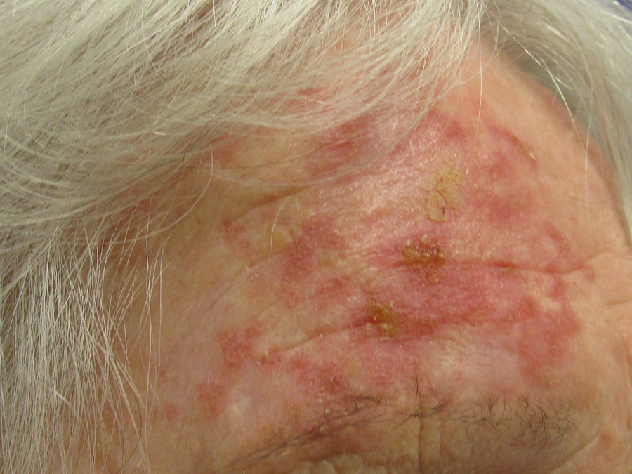
Above: Mild fluorouracil reaction

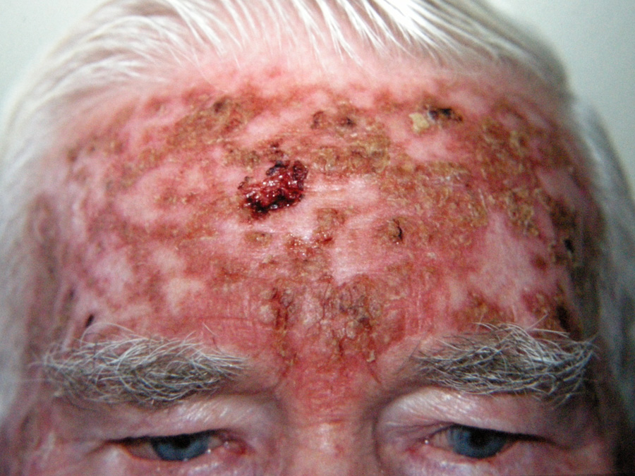
Above: Severe fluorouracil reaction

For further information and images of expected and extreme reactions to fluorouracil treatment, see: www.dermnetnz.org/topics/5-fluorouracil-cream/

Application is likely to cause erythema, erosion, excoriation/flaking, oedema and itching of affected skin. Painful erosions can occur on mucus membranes.


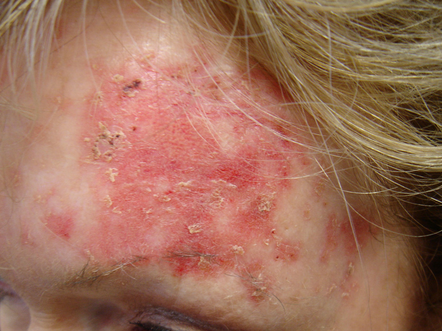
Above: Mild imiquimod reaction

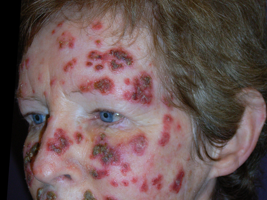
Above: Severe imiquimod reaction

For further information and images of expected and extreme reactions to imiquimod treatment, see: www.dermnetnz.org/topics/imiquimod/

Other adverse effects

Some patients may experience an intense reaction, including pain, burning, pruritus, rash, crusting, allergic contact dermatitis, hyperpigmentation, scarring, inflammation and photosensitivity

May result in secondary bacterial infection (via skin erosions)

May cause systemic symptoms including fever, nausea, diarrhoea, headache and mouth ulcers

May cause leukocytosis or leukopenia

Rarely associated with erythema multiforme

May cause localised hypopigmentation and hyperpigmentation, which has been reported to be permanent in some patients

May cause flu-like symptoms, e.g. fever, nausea, diarrhoea, headache and myalgia (symptoms are usually tolerated with paracetamol)

May cause reductions in haemoglobin, white blood cell count, absolute neutrophils and platelets

Rarely associated with erythema multiforme, Stevens-Johnson syndrome and cutaneous lupus erythematosus-like effect

Contraindications and cautions for use

Avoid use in pregnant or breastfeeding women

Avoid contact with eyes or mucus membranes, unaffected skin, broken skin or open wounds

Use cautiously in the perioral area or nasolabial fold, and on lesions below the knees as long-term ulceration may result from poor healing

Occlusion increases adverse effects

Avoid prolonged exposure to UV light as this will intensify the reaction to fluorouracil

Use with caution in pregnant or breastfeeding women

Use with caution in patients who are immunosupressed, have autoimmune disease or haematological abnormalities

Avoid contact with eyes or mucous membranes, unaffected skin, broken skin or open wounds

Not photosensitising, but prolonged exposure to UV light should still be avoided

Pre-treatment

When treating a hyperkeratotic lesion, pre-treatment with cryotherapy and/or a keratolytic such as 2% salicylic acid, urea cream or glycolic acid lotion will reduce scaling and therefore improve the absorption and efficacy of fluorouracil or imiquimod

Tretinoin cream may be used for two weeks prior to fluorouracil treatment to enhance the effect by peeling off the top layer of skin. This can also reduce the amount of time that fluorouracil needs to be used. N.B. Some patients with sensitive skin may not tolerate tretinoin.

Application

Wash skin with water and dry

Apply thinly with the tip of a finger (using gloves) or a cotton bud – wash hands immediately if not using a glove

The maximum area of skin that should be treated is 22 cm × 22cm

Hyperkeratotic lesions should be covered with an occlusive dressing to enhance absorption

A topical corticosteroid can be used for symptomatic relief if a severe reaction occurs

Apply thinly with the tip of a finger, rub into the skin (wash hands after application) and leave on for eight hours (typically overnight), then wash off any residue with soap and water

One sachet is sufficient to treat a 20 cm × 20 cm area of skin

It is not recommended to apply a topical corticosteroid to treat a reaction (unless severe) as this may affect the efficacy of imiquimod. If intolerable local adverse effects occur, the cream should be washed off.

If inflammation is extreme, consider less frequent application, a seven-day break from treatment or, if erosions are present, ceasing treatment

Dose for actinic keratoses

Apply once or twice daily, usually for three to four weeks

Monitor weekly for response

Treatment duration may need to be extended (up to eight weeks) if response is slow, e.g. for lesions on the trunk, lower limbs, hands and forearms, or shortened (one or two weeks) for lesions that are quick to respond, e.g. flat facial lesions

Apply two to three times a week for four to six weeks

Assess local response after three weeks and adjust treatment frequency if necessary

Review again after a four week treatment-free interval; treatment can be given for a further six to ten weeks if the lesion persists

Dose for superficial BCC

Apply twice daily, for six to 12 weeks

N.B. Fluorouracil is generally only used to treat small, very superficial BCC, and treatment is associated with a high rate of recurrence

Apply to the lesion, including a 1 cm surrounding margin, once daily on five days per week, for six weeks

Review after three weeks (or earlier if the patient reports an extreme reaction) and adjust frequency of application if necessary depending on response (e.g. reduce to three days per week)

Assess clinical outcome 6 – 12 weeks after treatment has ceased; treatment can be repeated for a further six to ten weeks if response has been inadequate, provided that there has been at least a four week break since the first treatment

N.B. best used for BCC <2 cm diameter

Dose for SCC in situ

Apply once or twice daily, for three to four weeks

Use an occlusive dressing to increase fluorouracil penetration if tissue reaction is minimal

Assess response; treatment can be continued for a further four weeks, or longer if necessary

Used off-label for this indication; regimen as for superficial BCC

Apply to the lesion, including a 1 cm surrounding margin, once daily on five days per week, for six weeks

Review after three weeks and adjust frequency of application if necessary depending on response (e.g. reduce to three days per week)

Assess clinical outcome 6 – 12 weeks after treatment has eased; treatment can be repeated for a further six to ten weeks if response has been inadequate, provided that there has been at least a four week break since the first treatment.

Post-treatment

Depending on the patients symptoms, petroleum jelly, an emollient or a mild topical corticosteroid can be used to assist healing.


Patient information on the use of fluorouracil and imiquimod is available from:
www.saferx.co.nz/Patient_info_fluorouracil.pdf
www.saferx.co.nz/imiquimod-patient-guide.pdf

Acknowledgement:

Thank you to Dr Amanda Oakley, Honorary Associate Professor and Dermatologist, Waikato District Health Board for expert review of this article.


References

  1. Newlands C, Currie R, Memon A, et al. Non-melanoma skin cancer: United Kingdom National Multidisciplinary Guidelines. J Laryngol Otol 2016;130:S125–32. http://dx.doi.org/10.1017/S0022215116000554
  2. Poulin Y, Lynde CW, Barber K, et al. Non-melanoma Skin Cancer in Canada Chapter 3: Management of Actinic Keratoses. J Cutan Med Surg 2015;19:227–38. http://dx.doi.org/10.1177/1203475415583414
  3. Krawtchenko N, Roewert-Huber J, Ulrich M, et al. A randomised study of topical 5% imiquimod vs. topical 5-fluorouracil vs. cryosurgery in immunocompetent patients with actinic keratoses: a comparison of clinical and histological outcomes including 1-year follow-up. Br J Dermatol 2007;157 Suppl 2:34–40. http://dx.doi.org/10.1111/j.1365-2133.2007.08271.x
  4. Zloty D, Guenther LC, Sapijaszko M, et al. Non-melanoma Skin Cancer in Canada Chapter 4: Management of Basal Cell Carcinoma. J Cutan Med Surg 2015;19:239–48. http://dx.doi.org/10.1177/1203475415586664
  5. New Zealand Formulary (NZF). NZF v54. 2016. Available from: www.nzf.org.nz (Accessed Dec, 2016)
  6. Bausch & Lomb (NZ) Ltd. New Zealand data sheet: ALDARA. 2015. Available from: www.medsafe.govt.nz/profs/datasheet/a/aldaracream.pdf
  7. Bausch & Lomb (NZ) Ltd. Data sheet: EFUDIX. 2016. Available from: www.medsafe.govt.nz/profs/datasheet/e/Efudixcr.pdf
  8. DermNet New Zealand. Skin cancer. Available from: www.dermnetnz.org/topics/skin-cancer/
  9. bpacnz. Managing non-melanoma skin cancer in priamry care. BPJ 2013; [Epub ahead of print]. Available from: www.bpac.org.nz (Accessed Jan, 2017)

Published: 20 January 2017 | Updated:

There are currently no comments for this article.

Please login to make a comment.

Made with by the bpacnz team

Partner links